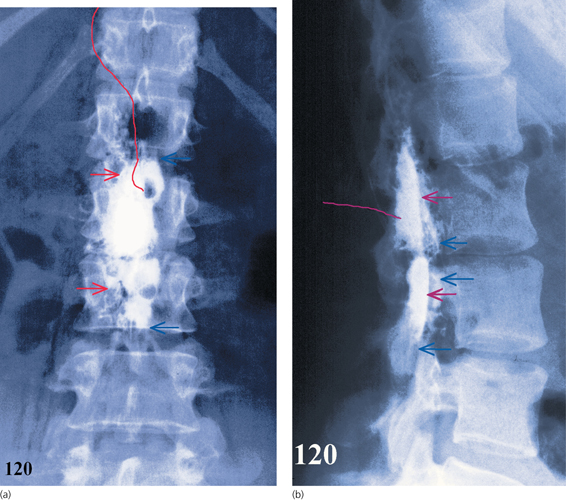
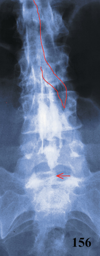
Figure 3.9
(a–d) Sequence of anteroposterior (AP) fluoroscopic images during contrast injection. (a) At 20s, a small central body of contrast appears (red arrows), with spill through the left L4–5 intervertebral foramen (blue arrow). (b) By 30s, the central body of contrast has widened and spread to L3 (lower red arrow), and lateral spread of contrast has appeared up to L2 on the right (upper red arrow). There is increased left L4–5 transforaminal nerve spill (blue arrow). (c) After 50s, the central body of contrast has again extended, while lateral spread has reached L1 on the left and T10 on the right (red arrows). The left L3–4 transforaminal spill has become more prominent and right L4–5 spill has appeared (blue arrows). (d) At 70s, the contrast has extended to left T12 and generally thickened in appearance. (e) The AP radiograph, in the same patient at 80s, shows the asymmetrical central body of contrast and the lateral channelling, which is a little patchy. The L3–4 and L4–5 transforaminal spill is arrowed in blue. (f) The lateral radiograph at 110s, showing a fairly typical spread of contrast, although it is patchy and attenuated in places. The L3–4 and 4–5 transforaminal nerve spill is arrowed in blue.
Epidural Anaesthesia / Hodder Arnold © 2012 C Collier