Figures
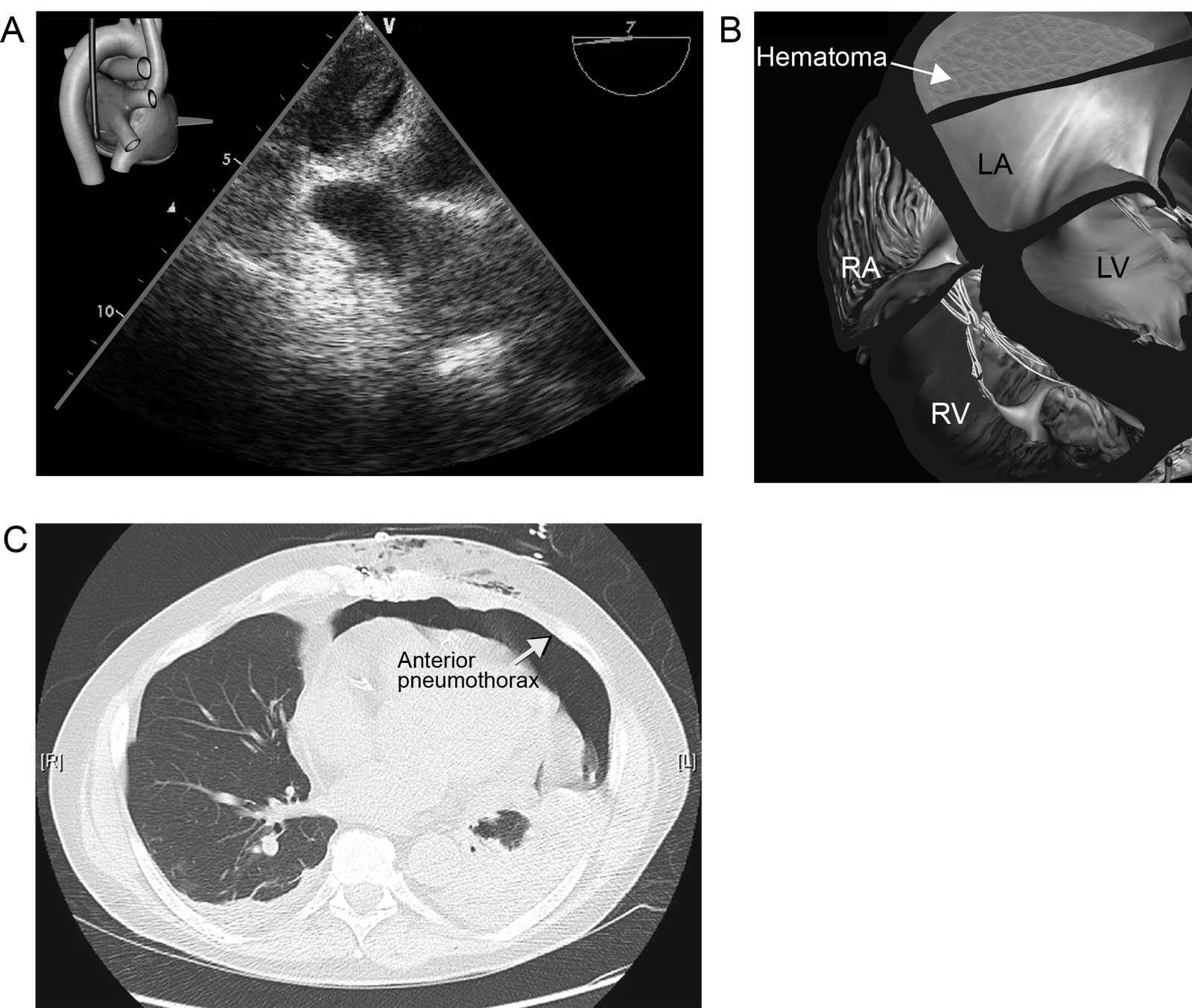
eFigure 12.4
Oblique sinus hematoma. (A, B) ME view at 7° shows a hematoma in the oblique sinus compressing the LA in an unstable patient after cardiac surgery. (C) The CT scan with an anterior pneumothorax (arrow) made a TTE approach impossible for the diagnosis of this condition. Abbreviations: CT, computed tomography; LA, left atrium; LV, left ventricle; ME, mid-esophageal; RA, right atrium; RV, right ventricle; TTE, transthoracic echocardiography.
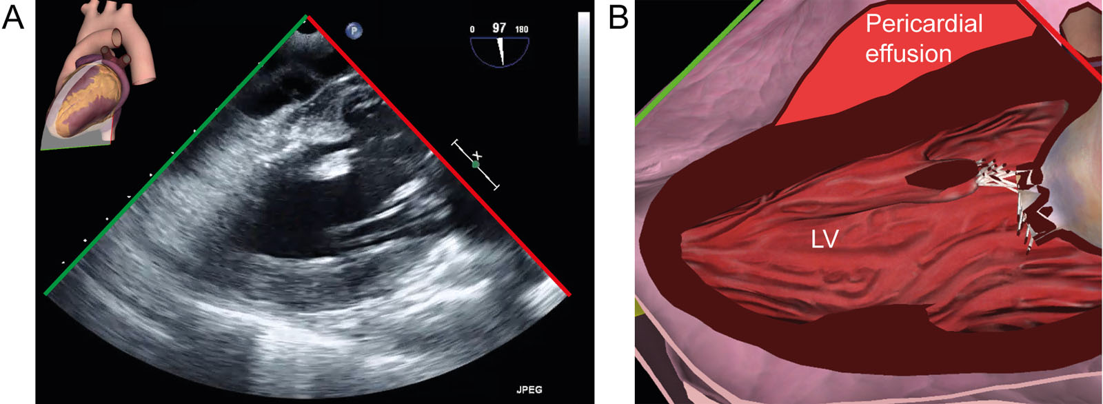
eFigure 12.5
Pericardial effusion. (A, B) TG 2C view shows a posterior pericardial effusion in an unstable 66-year-old man following cardiac arrest. Abbreviations: 2C, two-chamber; LV, left ventricle; TG, transgastric.
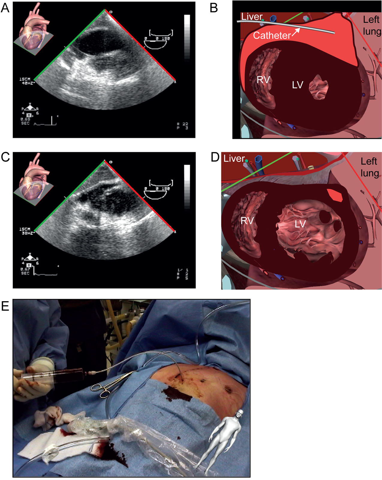
eFigure 12.13
Pericardial drainage. (A-E) These are TG SAX mid-papillary views (A, B) before and (C, D) after percutaneous pericardial drainage of a large (900 ml) pericardial effusion under TEE guidance (E) in the operating room. The guidewire appears behind the ventricles. Abbreviations: LV, left ventricle; RV, right ventricle; SAX, short-axis; TEE, transesophageal echocardiography; TG, transgastric. Source: Photo courtesy of Dr. Raymond Cartier.