Figures
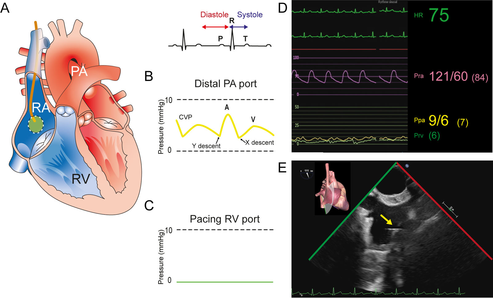
eFigure 13.2
TEE guided PAC insertion. (A) The PAC appears in the RA in this diagram. (B) The RA pressure or CVP waveform at the distal PAC tip, (C) without any signal in the pacing RV port. (D) The mean RA pressure is 7 mmHg. (E) ME bicaval view shows the PAC tip in the RA. Abbreviations: CVP, central venous pressure; HR, heart rate; ME, mid-esophageal; PA, pulmonary artery; PAC, pulmonary artery catheter; Ppa, pulmonary artery pressure; Pra, radial artery pressure; Prv, right ventricular pressure; RA, right atrium; RV, right ventricular; TEE, transesophageal echocardiography. Adapted from Raymond et al.103 (eFigure video 13.2 apply for eFigure 13.3 to 13.5).
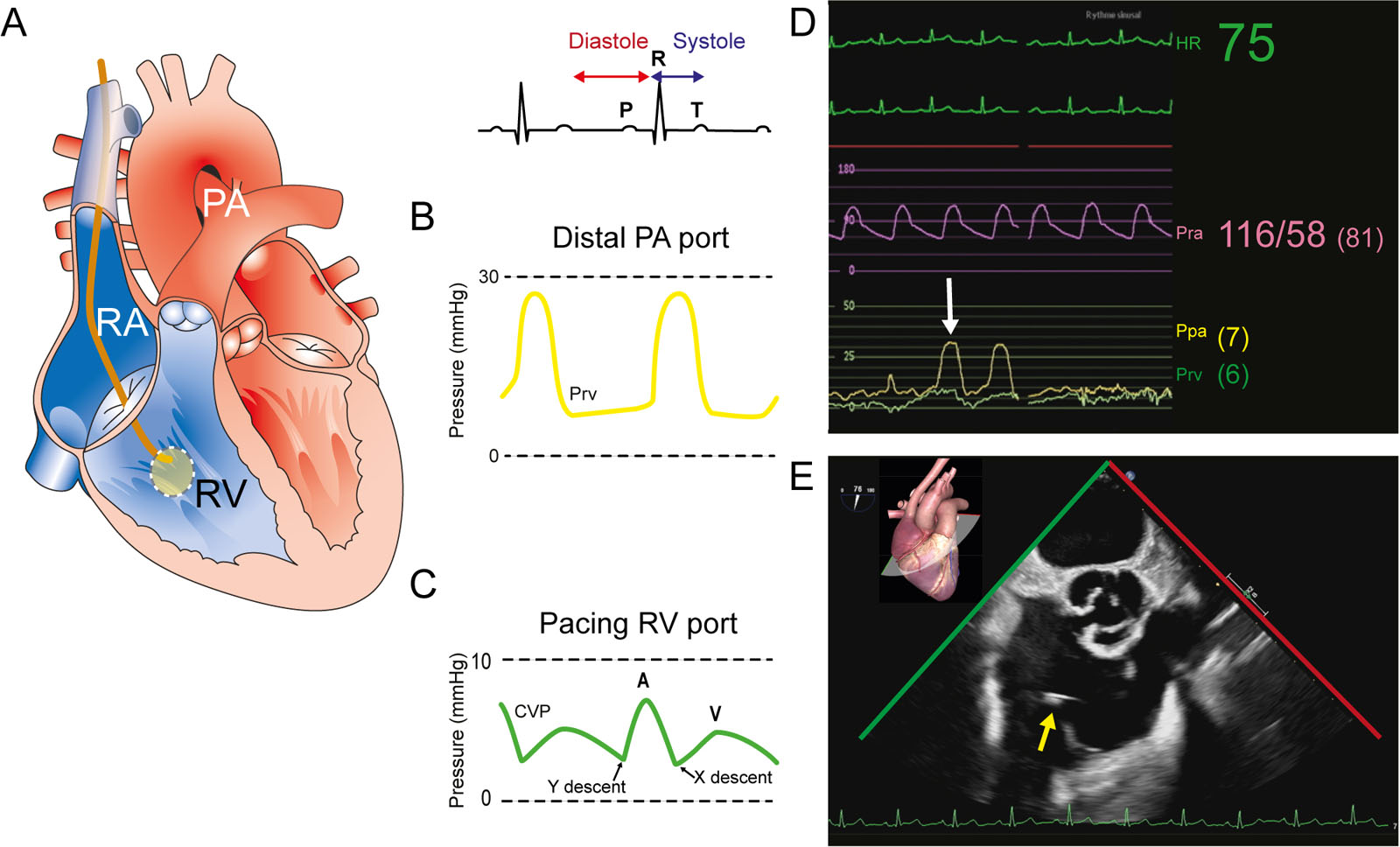
eFigure 13.3
TEE guided PAC insertion. (A) From Figure 13.2, the PAC advances into the RV, to show (B) the Prv waveform at the PAC distal tip and (C) the CVP waveform in the RV pacing port as seen in (D). (E) ME RV inflow-outflow view shows the PAC tip in the RV. Abbreviations: CVP, central venous pressure; HR, heart rate; ME, mid-esophageal; PA, pulmonary artery; PAC, pulmonary artery catheter; Ppa, pulmonary artery pressure; Pra, radial artery pressure; Prv, right ventricular pressure; RA, right atrium; RV, right ventricle; TEE, transesophageal echocardiography. Adapted from Raymond et al.103(eFigure video 13.2 apply for eFigure 13.3 to 13.5).
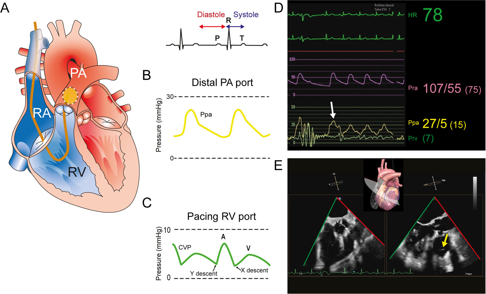
eFigure 13.4
TEE guided PAC insertion. (A) From Figure 13.3, the PAC advances into the PA, to show (B) the Ppa waveform at the PAC distal tip and (C) the CVP waveform in the RV pacing port, as seen in (D). (E) Biplane ME RV inflow-outflow view shows the PAC tip in the PA (arrow). Abbreviations: CVP, central venous pressure; HR, heart rate; ME, mid-esophageal; PA, pulmonary artery; PAC, pulmonary artery catheter; Ppa, pulmonary artery pressure; Pra, radial artery pressure; Prv, right ventricular pressure; RA, right atrium; RV, right ventricle; TEE, transesophageal echocardiography. Adapted from Raymond et al.103(eFigure video 13.2 apply for eFigure 13.3 to 13.5).
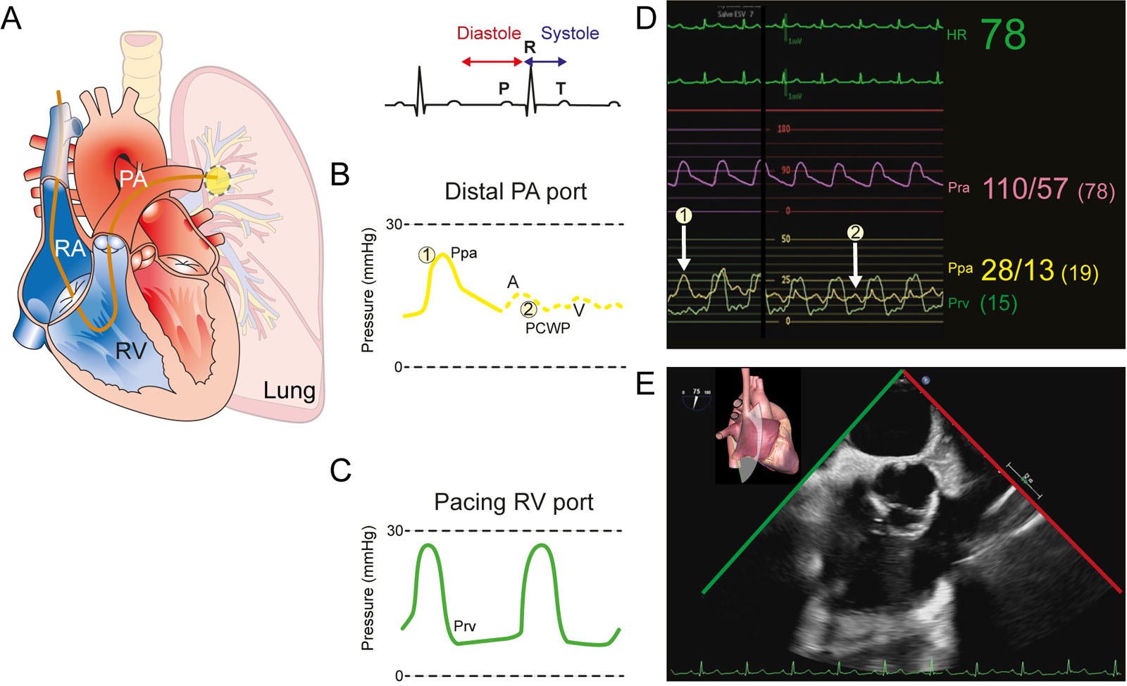
eFigure 13.5
TEE guided PAC insertion. (A) From Figure 13.4, the PAC advances further into the PA, to show the Ppa and PCWP waveforms at the PAC distal tip and (B) the Prv waveform in the RV pacing port with both, Ppa (arrow 1), Prv and PCWP (arrow 2), appearing in (C). (E) ME RV inflow-outflow view shows the PAC tip in the PA. Abbreviations: HR, heart rate; ME, mid-esophageal; PA, pulmonary artery; PAC, pulmonary artery catheter; PCWP, pulmonary capillary wedge pressure; Ppa, pulmonary artery pressure; Pra, radial artery pressure; Prv, right ventricular pressure; RA, right atrium; RV, right ventricle; TEE, transesophageal echocardiography. Adapted from Raymond et al.103(eFigure video 13.2 apply for eFigure 13.3 to 13.5).
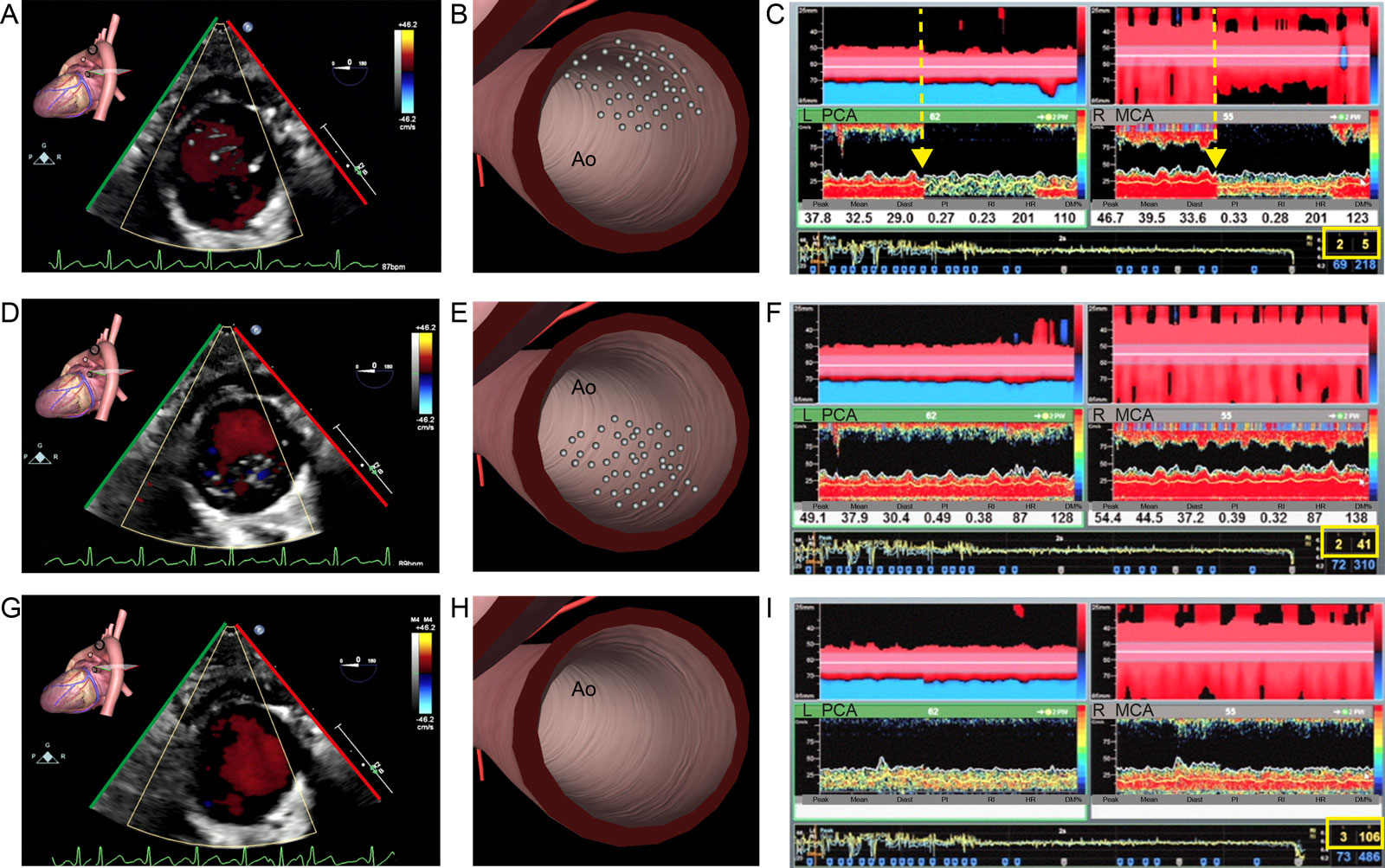
eFigure 13.7
Air emboli during initiation of CPB. (A-B) Desc Ao SAX view with CFI after initiation of CPB in minimally invasive cardiac surgery shows air in the Desc Ao with (C) a color change (arrow) in TCD monitoring. (D-I) Within a minute, the aortic air disappears in the Desc Ao SAX views with CFI and the TCD signal normalizes. The total number of high-intensity transient signals was 3 on the left PCA and 106 of the right MCA. Abbreviations: Ao, aorta; CFI, color flow imaging; CPB, cardiopulmonary bypass; Desc Ao, descending aorta; Diast. Diastole; DM%, % difference of mean velocity; HR, heart rate; L, left; MCA, middle cerebral artery; PCA, posterior cerebral artery; PI, pulsatility index; R, right; RI, resistance index; SAX, short-axis; TCD, transcranial Doppler.
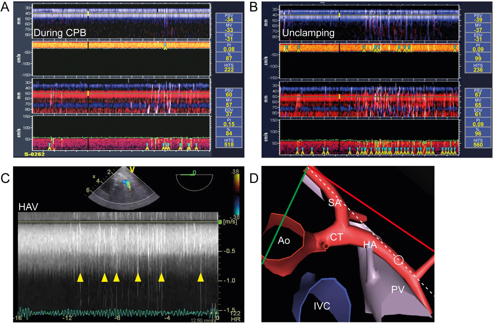
eFigure 13.8
Emboli during CPB. This is a 60-year-old man with ischemic cardiomyopathy and a LVEF < 30%. (A) TCD shows HITS (small A in yellow) during CPB after injecting 10 ml in the central line. (B) Upon release of the aortic cross clamp, additional HITS (yellow triangles) appeared simultaneously with (C, D) the abnormal HAV PWD signal. The HITS preceded VF. Abbreviations: %, percentage of mean velocity in relation to the baseline value; Ao aorta; CPB, cardiopulmonary bypass; CT, celiac trunk; EDV, end-diastolic velocity; HA, hepatic artery; HAV, hepatic artery velocities; HITS, high-intensity transient signals; IVC, inferior vena cava; LVEF, left ventricular ejection fraction; MV, mean velocity; PI, pulsatility index; PSV, peak systolic velocity; PV, portal vein; PWD, pulsed-wave Doppler; SA, splenic artery; TCD, transcranial Doppler; VF, ventricular fibrillation.
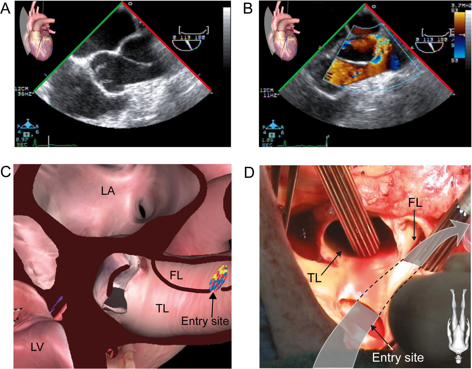
eFigure 13.12
Iatrogenic aortic dissection. (A–C) ME AoV LAX views without and with CFI demonstrate an intimal flap extending to the sinotubular junction with (B, C) flow from the true to the false lumen through an entry site. The aortic dissection resulted most likely from a tear in the posterior Ao wall at the site of previous aortic cannula insertion. (D) Surgical photo of the aorta shows the tear and the entry site in the aorta. Abbreviations: Ao, aorta; AoV, aortic valve; CFI, color flow imaging; FL, true lumen; LA, left atrium; LAX, long-axis; LV, left ventricle; ME, mid-esophageal; TL, true lumen. Source: Photo D courtesy of Dr. Michel Carrier.
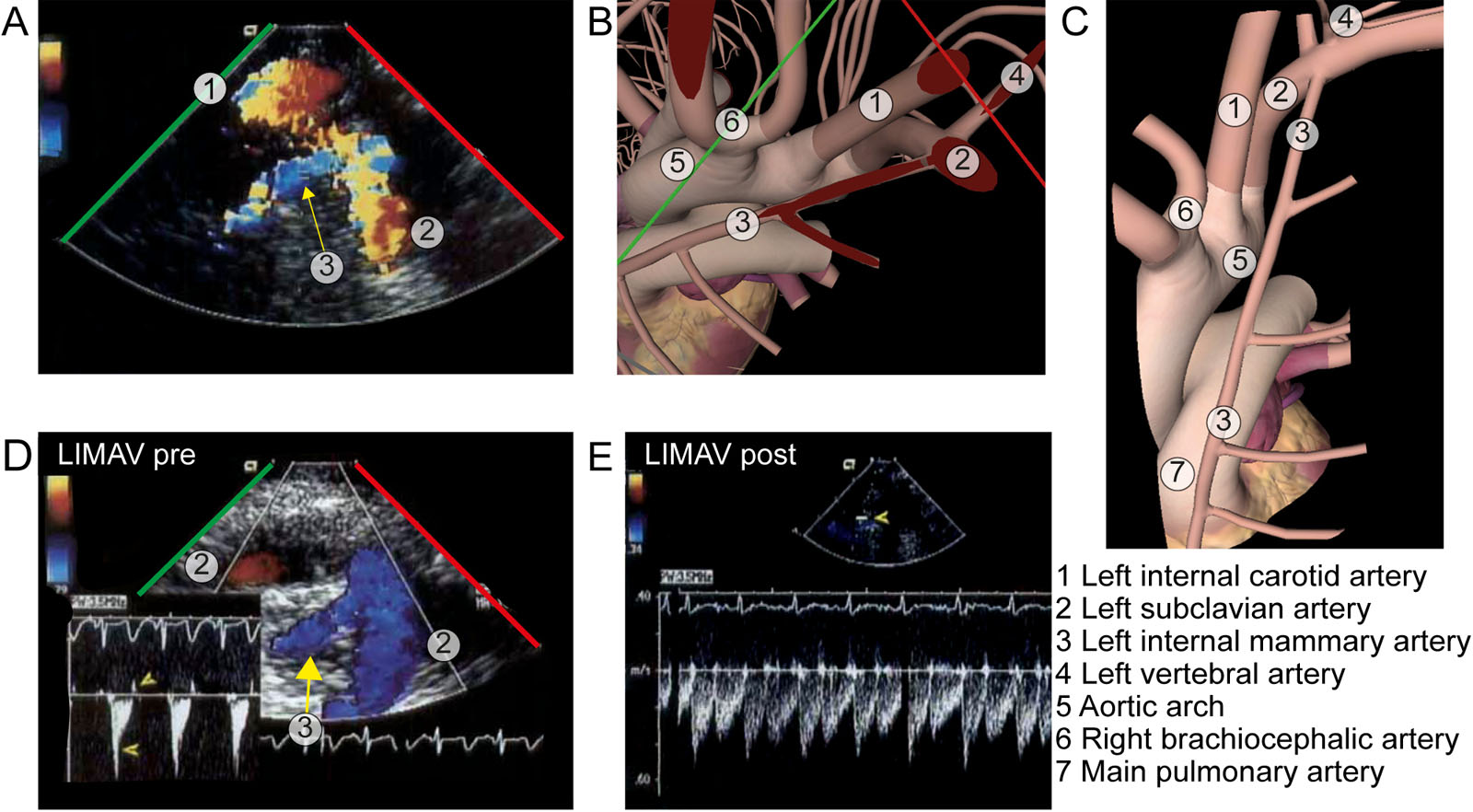
eFigure 13.23
LIMA view. (A-C) UE view with CFI and diagrams showing the LIMA arising from the left subclavian artery. (D, E) PWD interrogation of the LIMA before and after coronary revascularisation show increases in diastolic velocity after anastomosis of the LIMA to the LAD. Abbreviations: CFI, color flow imaging; LAD, left anterior descending artery; LIMA, left internal mammary artery; LIMAV, left internal mammary artery velocity; PWD, pulsed-wave Doppler; UE, upper esophageal. Adapted from Nanda et al.139
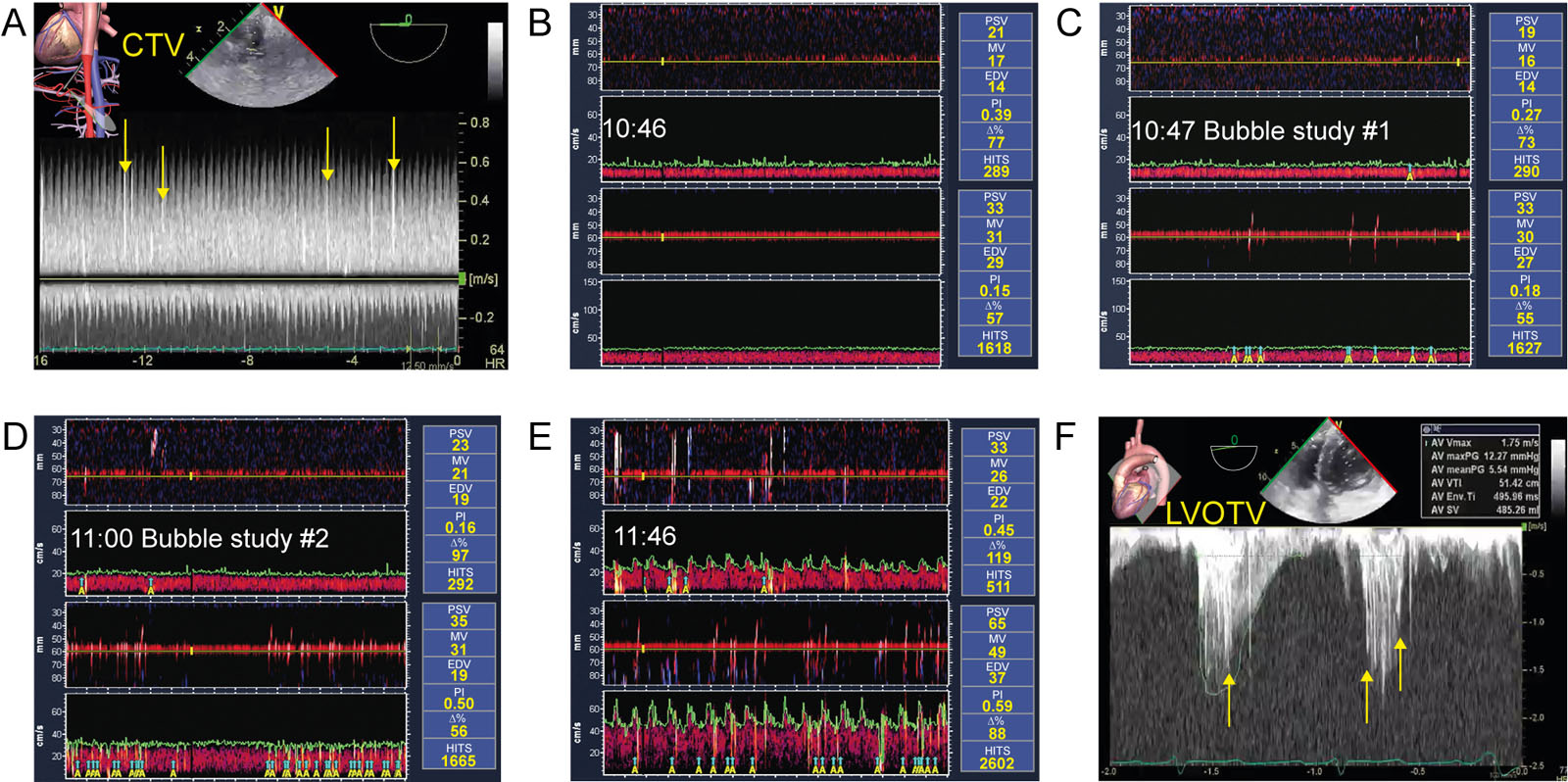
eFigure 13.25
Air emboli during and after CPB. (A) PWD interrogation of the celiac trunk during CPB shows HITS as white irregular signals. (B-E) TCD monitoring during CPB. A significant amount of HITS on the right (289) and on the left (1618) were present at the first bubble study. Air appeared after and was detected mostly on the left side. A total of 511 and 2602 HITS were present on the right and the left middle cerebral artery at the end of the procedure. The number of HITS correlates with post-op organ dysfunction.58 (F) DTG CWD velocity across the LVOT. Note again the HITS signal or white linear signals (arrows) as air was present in the LV after CPB. Abbreviations: CPB, cardiopulmonary bypass; CTV, celiac trunk velocity; CWD, continuous wave Doppler; DTG, deep transgastric; EDV, end-diastolic velocity; HITS, high intensity transient signals; LV; left ventricle, LVOT, left ventricular outflow tract; LVOTV, left ventricular outflow tract velocity; MV, mean velocity; PI, pulsatility index; PSV, peak systolic velocity; PWD, pulsed wave Doppler; TCD; transcranial Doppler. Adapted from Montrief et al.141
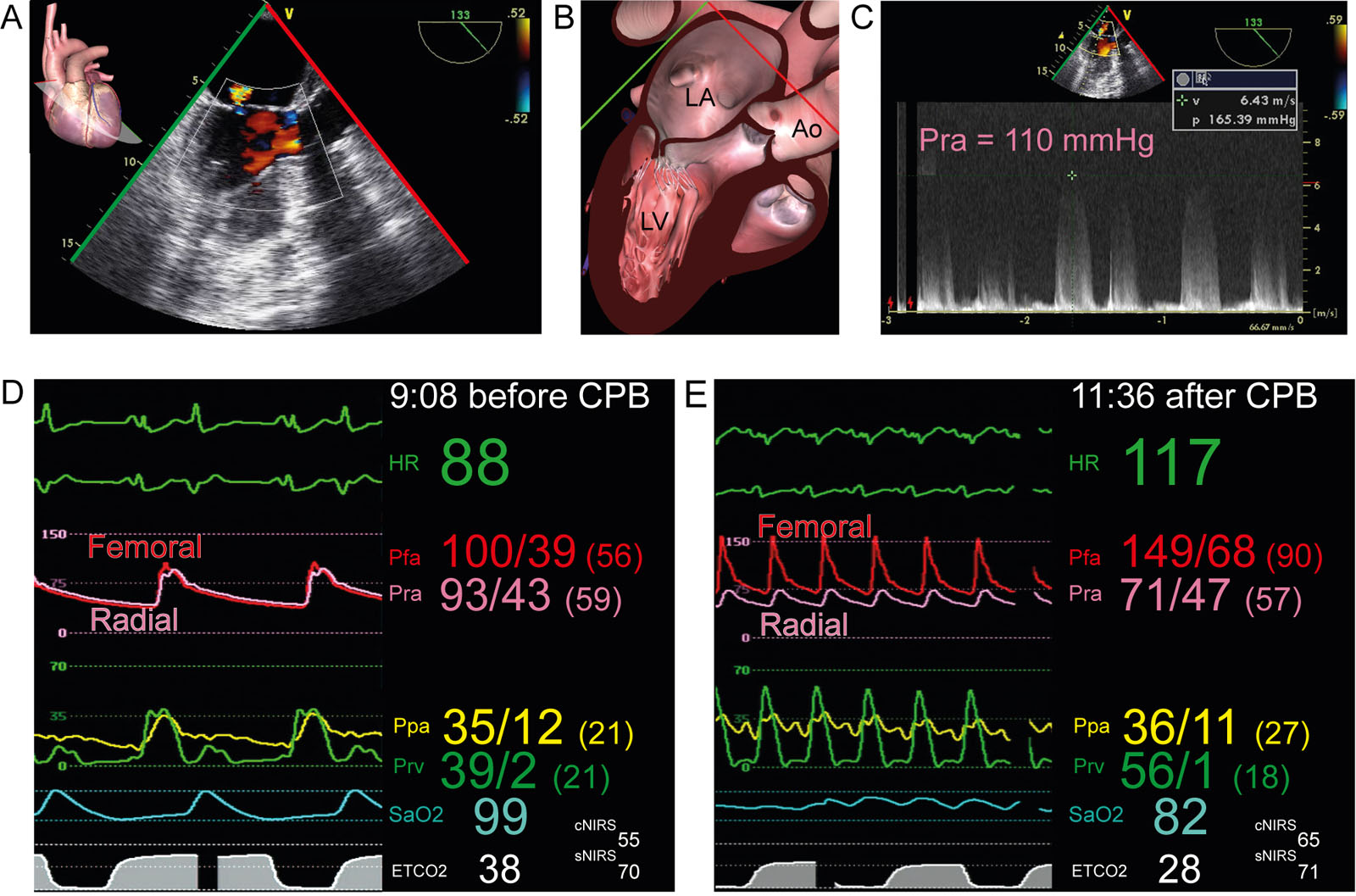
eFigure 13.26
Pseudo-radial artery hypotension. (A-C) This is a 54-year-old man hemodynamically unstable post-op after AoV endocarditis surgery. (A, B) The ME LAX view shows MR with (C) a CWD signal peak gradient of 165 mmHg, which differed from the systolic radial artery pressure of 110 mmHg. Insertion of a femoral artery catheter confirmed the significant radial to femoral gradient, allowing weaning of vasoactive drugs. (D,E) Hemodynamic data before and after CPB in a 61-year-old man undergoing coronary revascularisation and MVR. Inotropic support was required for difficult separation from CPB. Note the significant systolic radial to femoral pressure gradient of 78 mmHg after CPB. Note that the post-CPB cerebral and somatic NIRS value were normal which was inconsistent with the radial artery pressure. Abbreviations: Ao, aorta; aortic valve; AoV, aortic valve; cNIRS, cerebral near-infrared spectroscopy; CPB, cardiopulmonary bypass; CWD, continuous wave Doppler; ETCO2, end-tidal carbon dioxide; HR, heart rate; LA, left atrium; LAX, long-axis; ME, mid-esophageal; MR, mitral regurgitation; MVR, mitral valve replacement; NIRS, near-infrared spectroscopy; P, pressure; Pfa, femoral artery pressure; Ppa, pulmonary artery pressure; Pra, radial artery pressure; Prv, right ventricular pressure; SaO2, arterial oxygen saturation; sNIRS, somatic near-infrared spectroscopy; V, velocity. See Denault at al.142
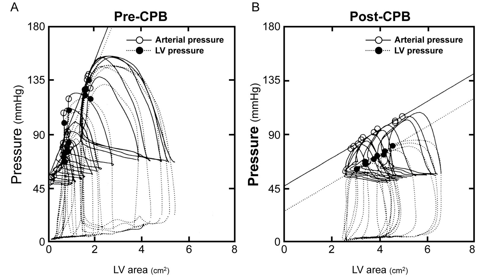
eFigure 13.27
Pressure-area loops. These are simultaneous arterial pressure-area loops (solid lines) and LV pressure-area loops (dashed lines) before (A) and after (B) CPB. The LV pressure end-systolic elastance (E′es) decreased from 49 to 12 mmHg/cm2 with a similar change in arterial E′es from 50 to 12 mmHg/cm2. The LV area is an estimate of LV volume. Abbreviations: CPB, cardiopulmonary bypass; LV, left ventricle. Reproduced with permission from Gorcsan et al.71
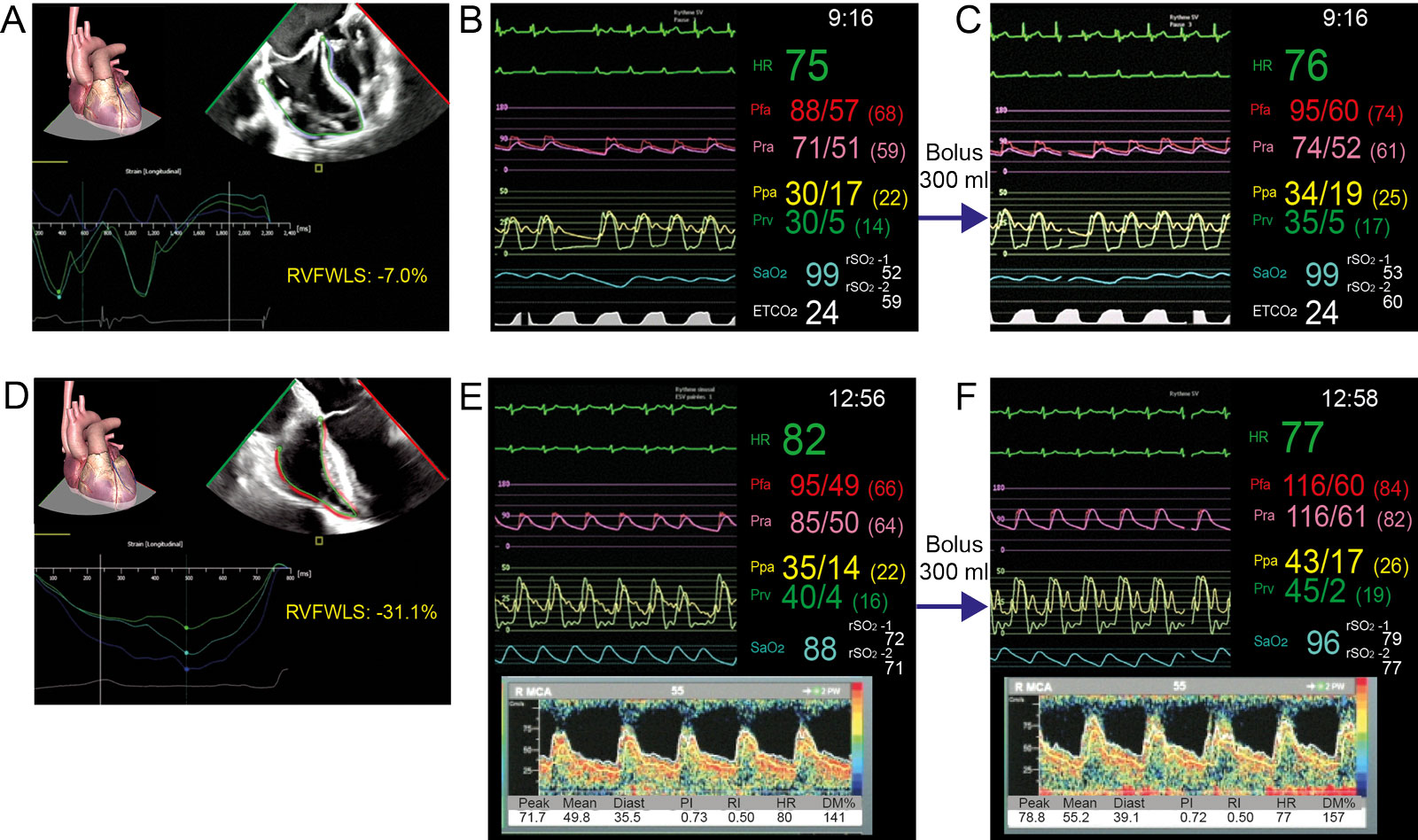
eFigure 13.28
RV strain and fluid responsiveness. (A-C) This is a 67-year-old man undergoing MV repair. The ME 4C view shows the RV FWSL strain of -7%. (B, C) These are the hemodynamic waveforms (B) before and (C) after a rapid 300 ml bolus of blood from the CPB reservoir. There are slight changes in Pra, Pfa, Ppa, Prv and rSO2 using NIRS, with no change in ETCO2. (D-F) This is a 67-year-old man undergoing AoV replacement and coronary revascularization. (D) The ME 4C view shows the RV FWSL of -30.1%. (E, F) These are the hemodynamic waveforms (E) before and (F) after a rapid 300 ml bolus of blood from the CPB reservoir. Here, there are significant changes in Pra, Pfa, Ppa, Prv, rSO2 and transcranial velocities. Abbreviations: 4C, four-chamber; AoV, aortic valve; CPB, cardiopulmonary bypass; Diast, diastolic; DM%, % velocity in relation to baseline; ETCO2, end-tidal carbon dioxide; HR, heart rate; ME, mid-esophageal; MV, mitral valve; NIRS, near infrared spectroscopy; Pfa, femoral artery pressure; PI, pulsatility index; Ppa, pulmonary artery pressure; Pra, radial artery pressure; Prv, right ventricular pressure; RI, resistance index; rSO2, regional oxygen saturation; RV, right ventricular; RVFWLS, right ventricular free wall longitudinal strain; SaO2, arterial oxygen saturation.
eFigure 13.38
Segmental dyskinesia. (A-D) ME LAX view during circumflex grafting shows isolated dyskinesia comprising paradoxical inward movement during (A, B) diastole and not systole (C, D) of the basal anterior septum. This occurs from verticalization and rotation of the heart while the Asc Ao assumes a fixed position and not LCx ischemia, which does not supply this septal region. The vertical cardiac displacement also elongates and enlarges the LA. Abbreviations: Ao, aorta; Asc Ao, ascending aorta; LA, left atrium; LAX, long-axis; LCx, left circumflex; LV, left ventricle; ME, mid-esophageal.
eFigure 13.39
Gas emboli. This patient is undergoing obtuse marginal artery off-pump bypass. (A) ME RV inflow-outflow view shows a massive gas embolism at the RCC obstructing the RCA ostium, causing severe RV dysfunction. (B) ME LAX view shows gaseous particles in the Asc Ao, LV and compacted in the LV apex (arrow). The LV apex assumes the uppermost position of the heart after cardiac displacement and verticalization. Abbreviations: Ao, aorta; AoV, aortic valve; Asc Ao, ascending aorta; LA, left atrium; LAX, long-axis; LV, left ventricle; ME, mid-esophageal; PA, pulmonary artery; RA, right atrium; RCA, right coronary artery; RCC, right coronary cusp; RV, right ventricular; TV, tricuspid valve.