Figures
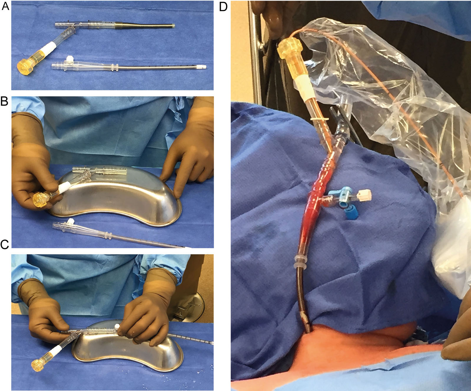
eFigure 20.19
Endovascular vena cavae occlusion technique. (A) Arterial Y-shaped cannula and arterial cannula used as a venous cannula. (B) The anesthesiologist cuts the arterial cannula portion and keeps the EndoReturn hemostasis valve portion. (C) The modified venous cannula for right internal jugular vein cannulation with the EndoReturn hemostatic valve portion connected to the arterial cannula. (D) CODA balloon catheter introduced into the modified venous cannula. The CODA catheter inserts into a sterile sheath. Adapted from Yamani et al.52
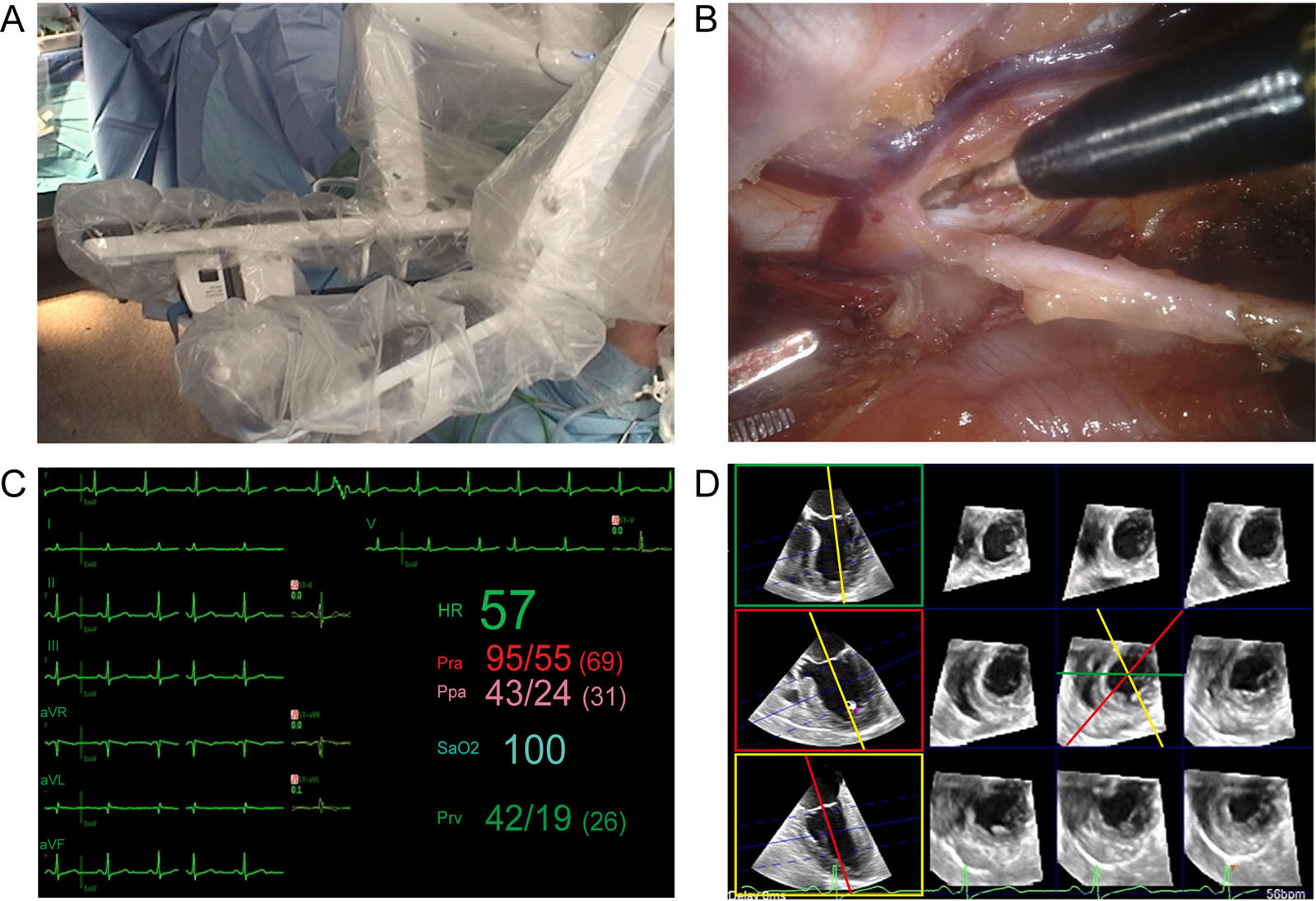
eFigure 20.21
Robotic surgery monitoring. (A,B) External and internal view of the robotic system used to perform coronary revascularization. Lung isolation allows LIMA dissection. (C,D) Electrocardiographic (7 lead) and 3D multi-slice reconstruction monitoring of myocardial ischemia. Abbreviations: 3D, three-dimensional; HR, heart rate; LIMA, left internal mammary artery; Ppa, pulmonary artery pressure; Pra, radial arterial pressure; Prv, right ventricular pressure, SaO2, oxygen saturation.
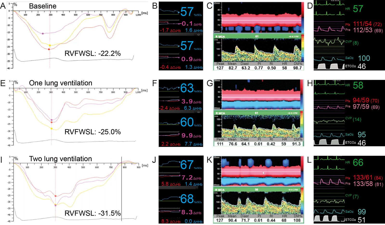
eFigure 20.24
RV strain and Anrep effect. A 68-year-old man is undergoing robotic revascularization with single lung ventilation. Compare the RVFWSL, second generation cerebral rSO2, TCD on the RMCA, and hemodynamic parameters at (A-D) baseline, (E-H) during one lung ventilation and (I-L) back on two lung ventilation. There is an increase in RVFWSL during one lung ventilation associated with a parallel increase in rSO2, ETCO2 and CVP, but transient reduction in TCD and both Pfa and Pra. The combination of a rise in carbon dioxide and an Anrep effect can explain the increase in strain and in rSO2 values. Abbreviations: DcHbi, change in total hemoglobin index; DHhbi, change in deoxygenated hemoglobin index; DO2Hbi, change in oxygenated hemoglobin index; CVP, central venous pressure; Diast, diastolic; DM%, delta mean or % compared to baseline mean velocity; ETCO2, end-tidal carbon dioxide; HR, heart rate; Pfa, femoral arterial pressure;PI, pulsatility index; Pra, radial arterial pressure; RI, resistance index; RMCA, right middle cerebral artery; rSO2, regional oxygen saturation; RV, right ventricle; RVFWSL, right ventricular free wall strain longitudinal; SaO2, oxygen saturation; TCD, transcranial Doppler.