Figures
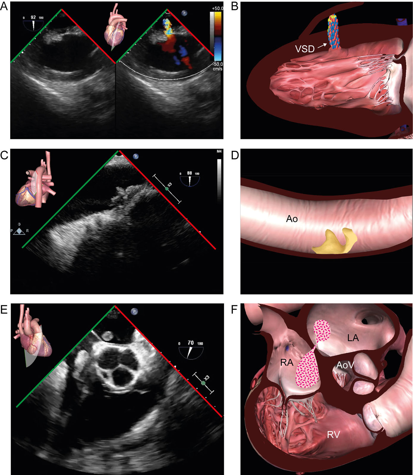
eFigure 21.1
Assist devices contraindications. These are some contraindications to the insertion of assist devices, including (A,B) a VSD seen in the TG 2C view, (C,D) grade 5 aortic atherosclerosis and a mobile plaque seen in the LAX thoracic aortic view and (E, F) thrombus across a PFO appearing in the ME RV inflow-outflow view. Abbreviations: 2C, two-chamber; Ao, aorta; AoV, aortic valve; LA, left atrium; LAX, long-axis; ME, mid-esophageal; PFO, patent foramen ovale; RA, right atrium; RV, right ventricle; TG, transgastric; VSD, ventricular septal defect.
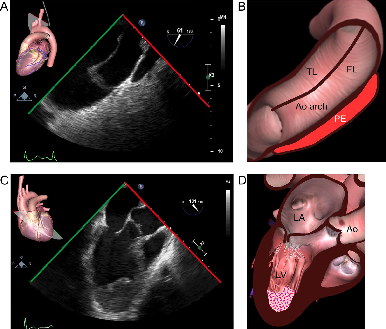
eFigure 21.7
Impella® device contraindications. Some contraindications to using the Impella® device include (A,B) aortic dissection as seen in this UE view of the aorta and (C,D) LV apical thrombus as appears in this ME LAX view. Abbreviations: Ao, aorta; FL, false lumen; LA, left atrium; LAX, long-axis; LV, left ventricle; ME, mid-esophageal; PE, pericardial effusion; TL, true lumen; UE, upper esophageal.
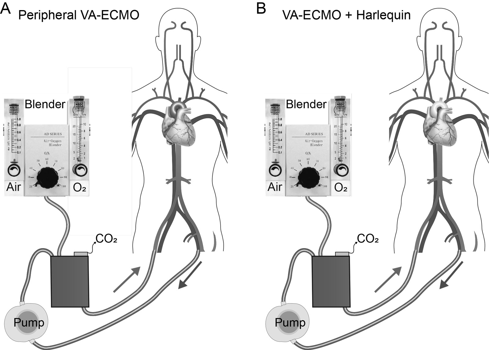
eFigure 21.17
Peripheral V-A ECMO. (A) Normal gas exchange. (B) Differential gas exchange. Red blood: reinfused oxygenated blood from the ECMO circuit. Blue blood: venous blood drained by ECMO circuit. Purple blood: relatively deoxygenated blood ejected from the LV in the setting of impaired native lung gas exchange leading to a Harlequin syndrome where the face and upper extremity are blue and the legs are red. Abbreviations: ECMO, extracorporeal membrane oxygenator; LV, left ventricular; V-A, veno-arterial. Adapted from Asija et al.37
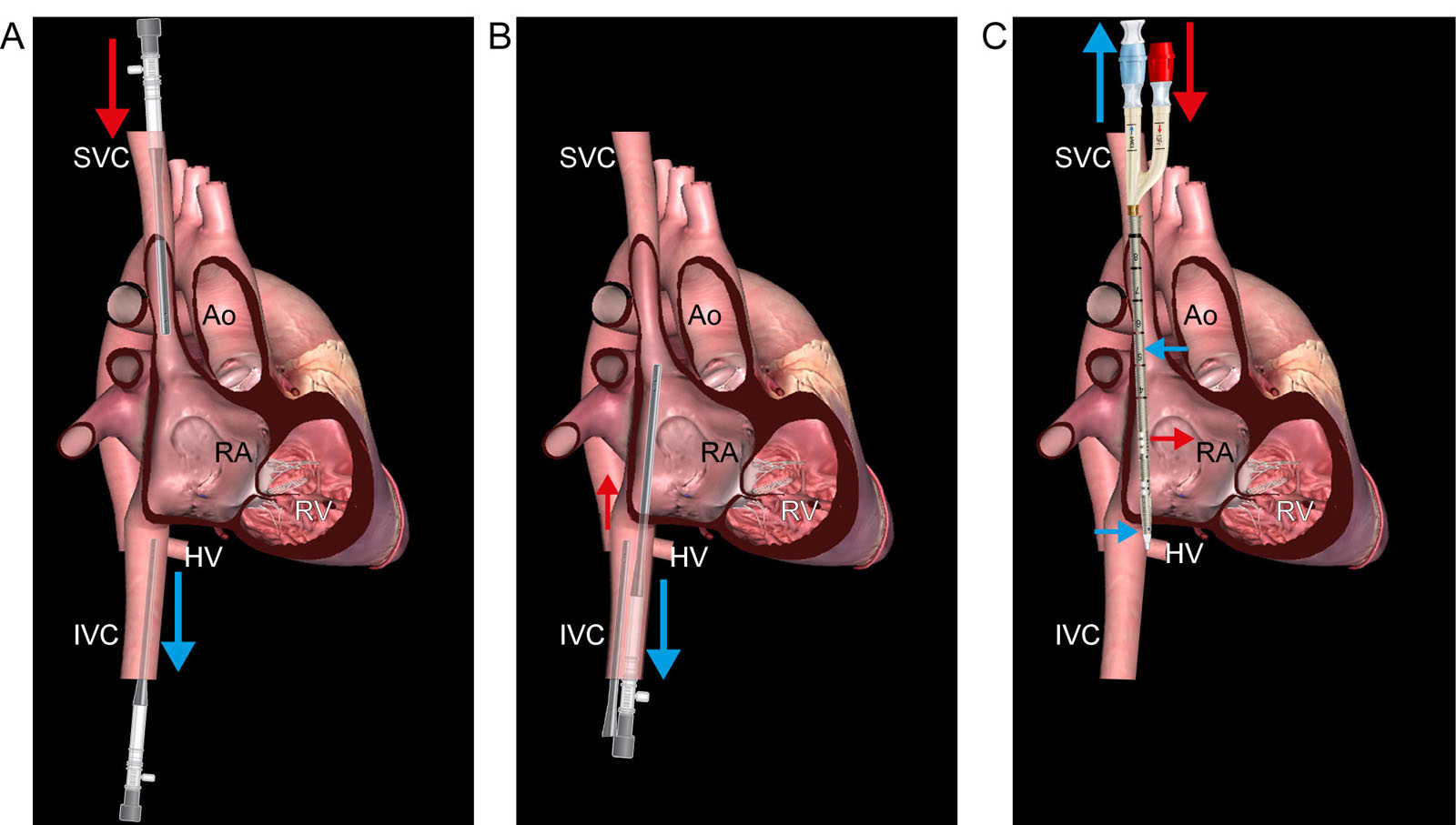
eFigure 21.18
V-V ECMO cannulation techniques. These diagrams show a few different options for V-V ECMO cannulation, including (A) femoral-jugular, (B) femoral-femoral and (C) dual-lumen cannula. In femoral-femoral cannulation, a distance ≥ 5 cm must separate the tips of the drainage and reinfusion/return cannula to avoid recirculation. Abbreviations: Ao, aorta; ECMO, extra-corporeal membrane oxygenation; HV, hepatic vein; IVC, inferior vena cava; RA, right atrium; RV, right ventricle; SVC, superior vena cava; V-V, veno-venous. Adapted from Banfi43 and Burrel et al.96
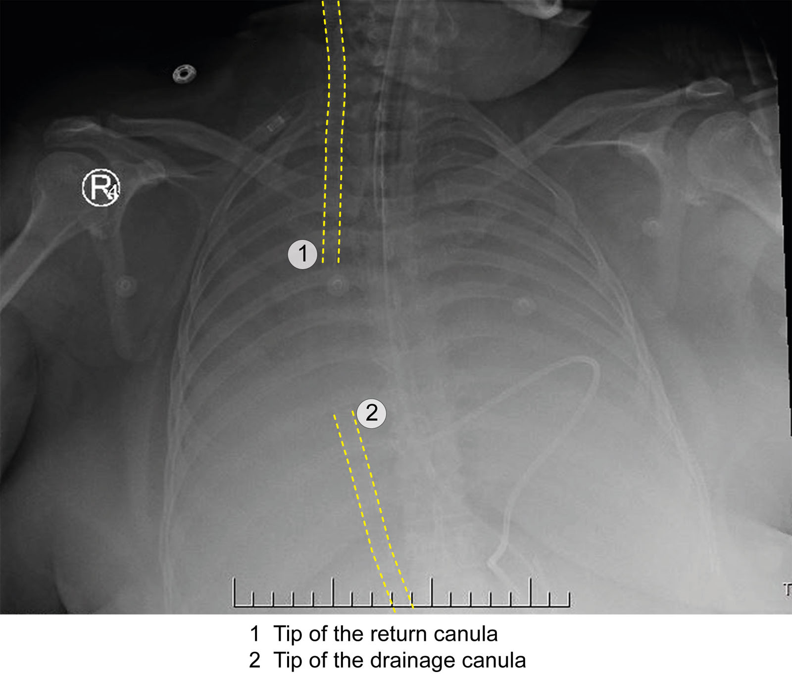
eFigure 21.19
V-V ECMO cannulae position. This CXR shows the multistage access cannula tip positioned at the RA/SVC junction, whereas the return cannula is ≥ 5cm above the proximal drainage hole. Abbreviations: CXR, chest x-ray; ECMO, extracorporeal membrane oxygenation; RA, right atrium; SVC, superior vena cava; V-V, veno-venous. Adapted from the The Alfred ICU (https://www.alfredicu.org.au/) and the Intensive blog (https://intensiveblog.com/ecmo/).
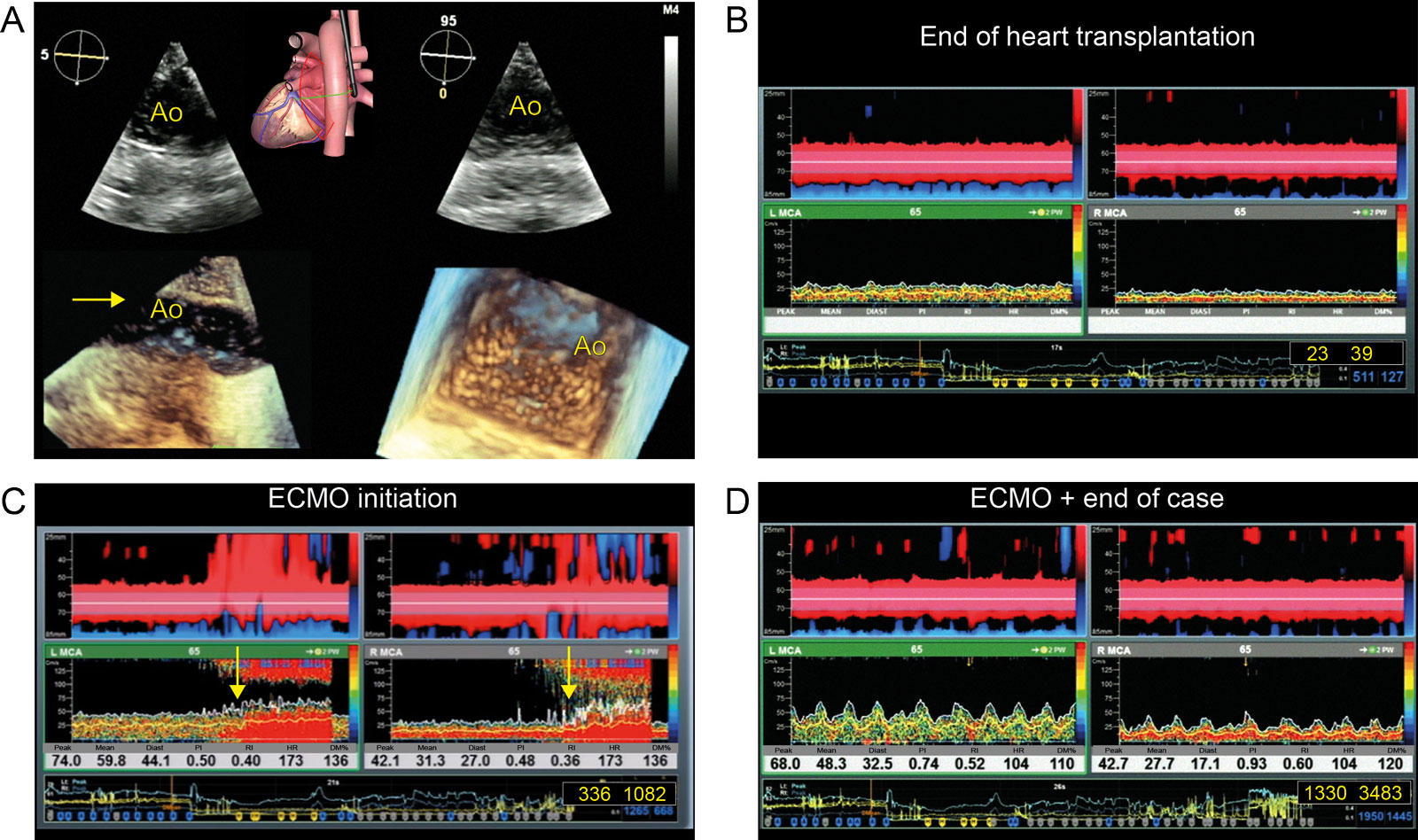
eFigure 21.24
Brain monitoring during ECMO. This is a patient undergoing heart transplantation who required ECMO. (A) Descending aorta biplane and 3D views show air (arrows) in the aortic lumen. (B-D) This was associated with the appearance of HITS in both the left and right cerebral territories (yellow arrows). Following ECMO initiation, the total number of HITS increased significantly on the right from 23 to 1330 and on the left from 39 to 348. Abbreviations: 3D, three-dimensional; Ao, aorta; Diast, diastole; DM%, % percentage of mean velocity changes or delta in relation to the baseline value; ECMO, extra-corporeal membrane oxygenation; HITS, high-intensity transient signals; HR, heart rate MCA, right middle cerebral artery; PI, pulsatility index; RI, resistance index. Adapted from Noel et al. 54
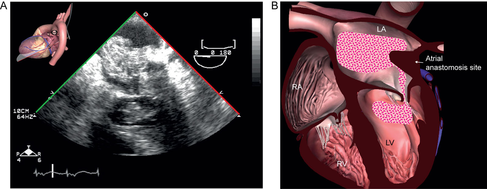
eFigure 21.25
ECMO complication. A 64-year-old man is on a centrifugal pump for cardiogenic shock after heart transplantation. (A, B) ME 4C views show an unexpected LA and LV thrombus attached to the atrial anastomosis site on the third day. Abbreviations: 4C, four chamber; ECMO, extracorporeal membrane oxygenation; LA, left atrium; LV, left ventricle; ME, mid-esophageal; RA, right atrium; RV, right ventricle.
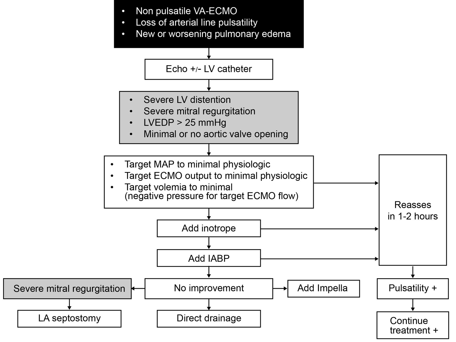
eFigure 21.26
LV venting. This algorithm describes LV venting diagnosis and therapeutic options in patients with V-A ECMO. Abbreviations: ECMO, extracorporeal membrane oxygenation; IABP, intra-aortic balloon pump; LA, left atrial; LV, left ventricular; LVEDP, left ventricular end-diastolic pressure; MAP, mean arterial pressure; VA, veno-arterial. Adapted from Ricarte Bratti et al.98
Videos
Chapter 21 Fig01A
Chapter 21 Fig01C
Chapter 21 Fig01E
Chapter 21 Fig03A
Chapter 21 Fig03CE
Chapter 21 Fig03E
Chapter 21 Fig04A
Chapter 21 Fig04D
Chapter 21 Fig05A
Chapter 21 Fig05D
Chapter 21 Fig06A
Chapter 21 Fig07A
Chapter 21 Fig07C
Chapter 21 Fig08A
Chapter 21 Fig08C
Chapter 21 Fig08D
Chapter 21 Fig08E
Chapter 21 Fig09A
Chapter 21 Fig09C
Chapter 21 Fig11A
Chapter 21 Fig11C
Chapter 21 Fig11D
Chapter 21 Fig11F
Chapter 21 Fig11G
Chapter 21 Fig11I
Chapter 21 Fig12A
Chapter 21 Fig12E
Chapter 21 Fig13A
Chapter 21 Fig13C
Chapter 21 Fig14A
Chapter 21 Fig14C
Chapter 21 Fig14E
Chapter 21 Fig15A
Chapter 21 Fig15C
Chapter 21 Fig15D
Chapter 21 Fig20BD
Chapter 21 Fig21A
Chapter 21 Fig21E
Chapter 21 Fig22C
Chapter 21 Fig22D
Chapter 21 Fig22F
Chapter 21 Fig22G
Chapter 21 Fig23A
Chapter 21 Fig23D
Chapter 21 Fig24ABCD
Chapter 21 Fig25
Chapter 21 Fig28C
Chapter 21 Fig28F
Chapter 21 Fig29A
Chapter 21 Fig29C
Chapter 21 Fig30A
Chapter 21 Fig30CD
Tables
eTable 21.2 Percutaneous Ventricular Assist Devices
| Device | TandemHeartTM | Impella® |
| Manufacturer | CardiacAssist, Inc., Pittsburgh, Pennsylvania, U.S. | Abiomed, Inc., Danvers, Massachusetts, U.S. |
| Approval | CE Mark, FDA 2003 | CE Mark, Canada, FDA 2007 |
| Cannula | Venous drainage, arterial outflow | Arterial only |
| Pump | Centrifugal, extracorporeal | Axial flow, intracorporeal |
| Circuit | Venous transseptal catheter in LA aspirates blood into pump and delivers into femoral artery | Retrograde aortic catheter in LV through AoV aspirates blood from LV below valve and delivers into the ascending aorta above valve |
| Contraindications | RV failure (for left-sided devices), VSD, AR, PVD | Mechanical AoV, calcific aortic stenosis, PVD |
| Anticoagulation | ACT > 300 s during insertion, Maintain ACT > 200 or use anti-Xa measurement | Maintain ACT > 160 ms |
| Hemodynamic effects | ↑MAP, ↑CO, ↓afterload, ↓preload, ↑MVO2 | ↑MAP, ↑CO, ↓PCWP |
| Complications | Cannula trauma (cardiac perforation), thromboembolism, hypothermia, bleeding, infection | Device malfunction, bleeding, infection, thrombus |
| Abbreviations: ACT, activated clotting time; AoV, aortic valve; AR, aortic regurgitation; CE, European Community; CO, cardiac output; FDA, Food and Drug Administration; LA, left atrium; LV, left ventricle; MAP, mean arterial pressure; MVO2, myocardial oxygen consumption; PCWP, pulmonary capillary wedge pressure; PVD, peripheral vascular disease; RV, right ventricle; VSD, ventricular septal defect. Source: Adapted from Vegas et al .88 | ||