Figures
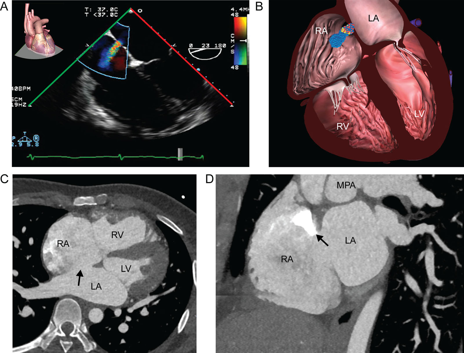
eFigure 26.2
ASD and RV dilatation. (A,B) ME 4C RV focused view with CFI shows dilatation of the RV and RA compared with the left-sided chambers during diastole. CFI shows left-to-right flow through the IAS from a secundum ASD. (C,D) CT scan view of an ASD. Note the IAS gap and the contrast entering the RA from the SVC (arrow at white zone) in a bi-atrial view.. Abbreviations: 4C, four-chamber; ASD, atrial septal defect; CFI, color flow imaging; CT, computed tomography; IAS, interatrial septum; LA, left atrium; LV, left ventricle; ME; mid-esophageal; MPA, main pulmonary artery; RA, right atrium; RV, right ventricle; SVC, superior vena cava.
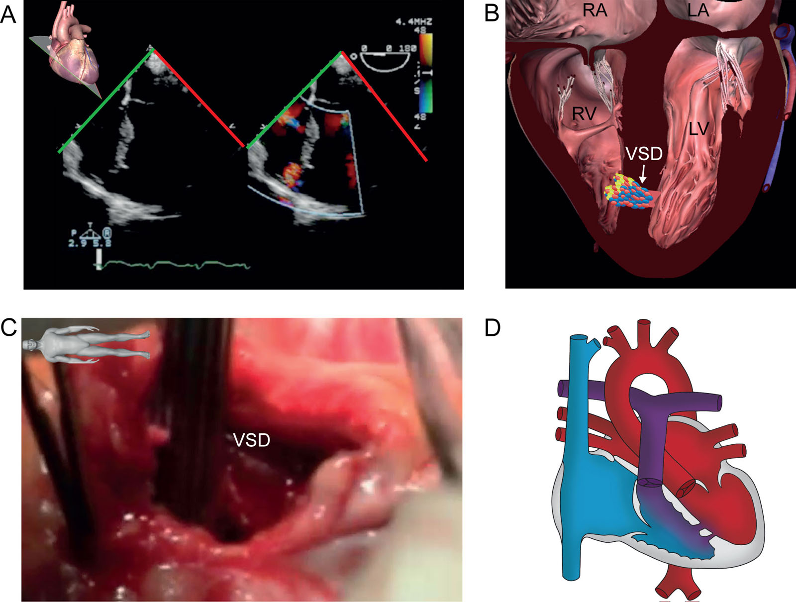
eFigure 26.18
Trabecular or muscular VSD. (A,B) ME color-compare 4C view with CFI shows a trabecular muscular VSD with left-to-right shunt confirmed by the (C) intraoperative findings. (D) The diagram illustrates blood flow related to the VSD shunt. Abbreviations: LA, left atrium; LV, left ventricle; ME, mid-esophageal; RA, right atrium; RV, right ventricle; VSD, ventricular septal defect.
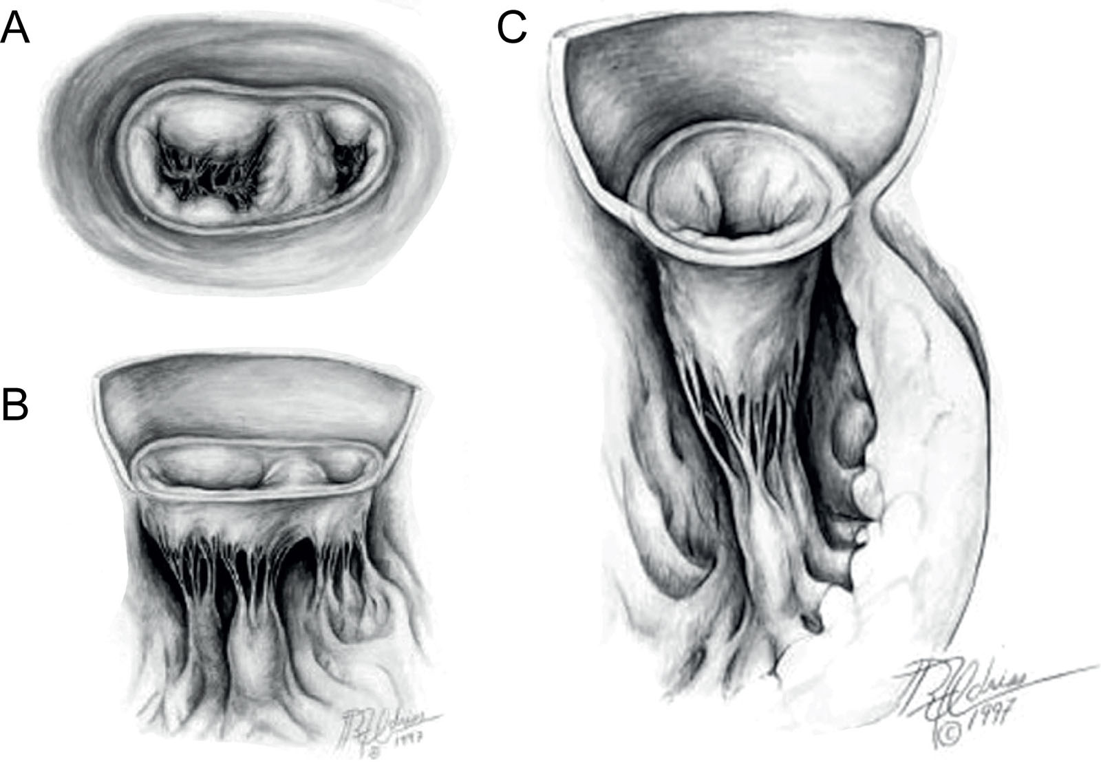
eFigure 26.31
31 Congenital MV abnormalities. (A,B) Double-orifice MV in these LA and cut-away atrioventricular diagrams, the smaller of the two orifices is in the right lateral position with variable stenosis. (C) Parachute MV in this cut-away atrioventricular diagram of a stenotic parachute MV demonstrates a single papillary muscle, often the postero-medial, or fused papillary muscles that usually arise from the inferior left ventricular wall. Abbreviations: LA, left atrial; MV, mitral valve. Adapted from Zias et al.40
Videos
Chapter 26 Fig02
Chapter 26 Fig04A
Chapter 26 Fig04E
Chapter 26 Fig05A
Chapter 26 Fig05C
Chapter 26 Fig05D
Chapter 26 Fig05F
Chapter 26 Fig06A
Chapter 26 Fig07A
Chapter 26 Fig08A
Chapter 26 Fig08D
Chapter 26 Fig08E
Chapter 26 Fig09A
Chapter 26 Fig10A
Chapter 26 Fig10C
Chapter 26 Fig11C
Chapter 26 Fig14A
Chapter 26 Fig14B
Chapter 26 Fig15A
Chapter 26 Fig16A
Chapter 26 Fig16B
Chapter 26 Fig16E
Chapter 26 Fig17A
Chapter 26 Fig17C
Chapter 26 Fig17E
Chapter 26 Fig18A
Chapter 26 Fig18C
Chapter 26 Fig19A
Chapter 26 Fig19D
Chapter 26 Fig21A
Chapter 26 Fig21C
Chapter 26 Fig22A
Chapter 26 Fig22C
Chapter 26 Fig22E
Chapter 26 Fig23A
Chapter 26 Fig24A
Chapter 26 Fig24B
Chapter 26 Fig24D
Chapter 26 Fig24F
Chapter 26 Fig25A
Chapter 26 Fig25B
Chapter 26 Fig25D
Chapter 26 Fig26A
Chapter 26 Fig26D
Chapter 26 Fig26G
Chapter 26 Fig27A
Chapter 26 Fig27C
Chapter 26 Fig27E
Chapter 26 Fig27F
Chapter 26 Fig28A
Chapter 26 Fig28B
Chapter 26 Fig28D
Chapter 26 Fig29A
Chapter 26 Fig29B
Chapter 26 Fig29D
Chapter 26 Fig30C
Chapter 26 Fig30E
Chapter 26 Fig30F
Chapter 26 Fig32A
Chapter 26 Fig32C
Chapter 26 Fig32E
Chapter 26 Fig32G
Chapter 26 Fig33AB
Chapter 26 Fig33FG
Chapter 26 Fig35D
Chapter 26 Fig36B
Chapter 26 Fig36C
Chapter 26 Fig36D
Chapter 26 Fig36E
Chapter 26 Fig37A
Chapter 26 Fig37D
Chapter 26 Fig37G
Chapter 26 Fig38A
Chapter 26 Fig38D
Chapter 26 Fig39A
Chapter 26 Fig39C
Chapter 26 Fig40A
Chapter 26 Fig40D
Chapter 26 Fig41A
Chapter 26 Fig42A
Chapter 26 Fig42C
Chapter 26 Fig42E
Chapter 26 Fig43A
Chapter 26 Fig45A
Chapter 26 Fig45D
Chapter 26 Fig46A
Chapter 26 Fig46D
Chapter 26 Fig47A
Chapter 26 Fig47D
Tables
eTable 26.2 Recommended TEE views to image congenital cardiac malformations
| Malformation | Views |
| Atrial septal defect (Figure 26.3) | ME 4C (0°-20°); ME AoV SAX (30°-60°); ME Bicaval (80°-110°) |
| Ventricular septal defect (Figure 26.13) | ME 4C (0°-20°); ME AoV SAX (30°-60°); ME AoV LAX (120°-160°); TG SAX (0°-30°) |
| Patent ductus arteriosus (Figure 26.23) | UE aortic short axis (90°); TG RV outflow (60°-90°) |
| AV canal defect or AVSD (Figure 26.20) | ME 4C (0°–20°); AoV LAX (120°–160°); TG basal SAX (0°–20°); TG LAX (90°–110°) |
| Ebstein’s anomaly (Figure 26.24) | ME 4C (0°–20°); ME RV outflow (60°–90°); TG LAX view of TV (60°–90°) |
| Pulmonary stenosis (See Figure 19.12) | UE RV outflow (60°–90°); TG RV outflow (60°–90°) |
| Tetralogy of Fallot (Figure 26.26) | ME 4C (0°–20°); ME AoV SAX (30°–60°); ME AoV LAX (120°–160°); TG RV outflow (60°–90°) |
| Cor triatriatum (Figure 26.30) | ME 4C (0°–20°); ME 2C (80°–100°) |
| Mitral valve stenosis (eFigure 26.31) | ME 4C (0°–20°); ME 2C (80°–100°); ME LAX (80°–100°); TG basal SAX (0°–25°); TG LAX (120°–160°) |
| Cleft mitral valve (Figure 26.22) | ME 4C (0°–20°); ME 2C (80°–100°); TG basal SAX (0°–20°) |
| Aortic valve stenosis (See Figure 14.31) | ME AoV SAX (30°–60°), ME AoV LAX (120°–160°); ME ascending aorta LAX (90°); TG LAX (90°–120°); DTG (0°–30°) |
| Subvalvular AS (See Figure 14.20) | ME 5C (0°–20°); TG LAX (90°–120°) |
| Supravalvular AS (See Figure 14.19) | ME AoV LAX (120°–160°); ME ascending aorta LAX (90°); DTG (0°–30°) |
| Coarctation of aorta (See Figure 23.45.) | UE aortic arch LAX (0°); UE aortic arch SAX (90°) |
| Truncus arteriosus (Figure 26.14) | ME 4C (0°–20°); ME AoV SAX (30°–60°); ME AoV LAX (120°–160°); TG RV outflow (60°–90°) |
| Transposition of the great arteries (Figure 26.34) | ME 4C (0°–20°); ME Bicaval (80°–110°); ME AoV SAX (30°–60°); ME AoV LAX (120°–160°); TG basal SAX (0°–20°); TG mid SAX (0°–20°) |
| Complex single ventricle (Figure 26.42) | ME 4C (0°–20°); ME Bicaval (80°–110°); ME AoV SAX (30°–60°); ME AoV LAX (120°–160°) TG RV outflow (60°–90°); TG basal SAX (0°–20°); TG Mid SAX (0°–20°) |
| Abbreviations: 2C, two-chamber; 4C, four chamber; 5C, five-chamber; AoV, aortic valve; AS, aortic stenosis; AVSD, endocardial cushion defect; DTG, deep transgastric; LAX. Long-axis; ME, mid-esophageal; RV, right ventricular; SAX, short-axis; SD, septal defect; TEE, transesophageal echocardiography; TG, transgastric; TV, tricuspid valve; UE, upper esophageal. | |