Figures
Videos
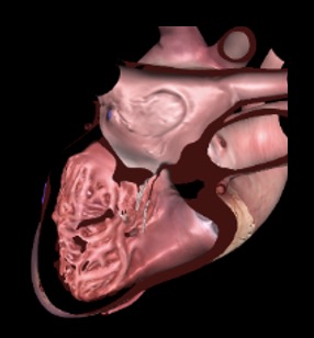
Chapter 29 Fig02B
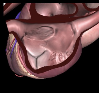
Chapter 29 Fig03A
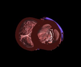
Chapter 29 Fig04AC
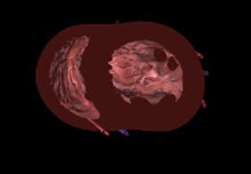
Chapter 29 Fig05A
Chapter 29 Fig06A
Chapter 29 Fig06B
Chapter 29 Fig06D
Chapter 29 Fig06G
Chapter 29 Fig09A
Chapter 29 Fig09C
Chapter 29 Fig09E
Chapter 29 Fig10A
Chapter 29 Fig10B
Tables
eTable 29.1 TEE views for non cardiac surgery and rescue TEE
TEE View |
View- |
Role |
Rescue & trauma TEE |
 |
ME 4C |
|
|
 |
ME MC |
|
|
 |
ME 2C view |
|
|
 |
ME LAX |
|
|
 |
ME AoV LAX |
|
|
 |
ME Ascending aorta SAX |
|
|
 |
ME AoV SAX |
|
|
 |
ME RV inflow-outflow |
|
|
 |
ME Modified bicaval TV |
|
|
 |
ME Bicaval |
|
|
 |
TG Basal SAX |
|
|
 |
TG Mid papillary SAX |
|
|
 |
DTG 5C |
|
|
 |
TG 2C LAX |
|
|
 |
Descending aorta SAX |
|
|
 |
Descending aorta LAX Angle: 90-100° |
|
|
Abbreviations: 2C, two-chamber; 4C, four-chamber; 5C, five-chamber; AoV, aortic valve; Asc Ao, ascending aorta; CFI, color flow imaging; CO, cardiac output; CPR, cardiopulmonary resuscitation; DTG, deep transgastric; IAS, interatrial septum; IVS, interventricular septum; LAA, left atrial appendage; LAX, long-axis; LV, left ventricle; LVOTO, left ventricular outflow tract obstruction; MC, mitral commissural; ME, mid-esophageal; MV, mitral valve; PA, pulmonary artery; PV, pulmonic valve; RVOTO, right ventricular outflow tract obstruction; SAX, short-axis; SV, splenic vein; SVC, superior vena cava; TEE, transesophageal echocardiography; TG, transgastric; TV, tricuspid valve; WMA, wall motion abnormalities. Adapted from Reeves94 , Fayad1, Giron-Arango et al.95 |
|||
eTable 29.4 Relative risk of vascular air embolism in common neurosurgical procedures
Common neurosurgical procedures |
Relative risk* |
|
Sitting position craniotomy |
High |
|
*Approximate expected reported incidences: high, > 25%; medium 5 to 25%; low, < 5%. Adapted from Mirski MA, et al.71 |
||