Figures
eFigure 3.11
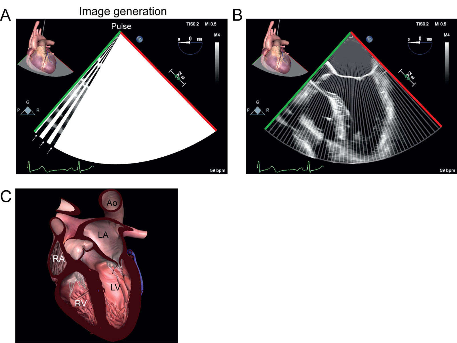
ME 5C view image generation. (A) A pulse creates each line in the image. (B, C) Summation of all the lines creates the final 2D image of the ME 5C view. Abbreviations: 2D, two dimensional; 5C, five-chamber; Ao, aorta; LA, left atrium; LV, left ventricle; ME, mid-esophageal; RA, right atrium; RV, right ventricle.
eFigure 3.11ME 5C view image generation. (A) A pulse creates each line in the image. (B, C) Summation of all the lines creates the final 2D image of the ME 5C view. Abbreviations: 2D, two dimensional; 5C, five-chamber; Ao, aorta; LA, left atrium; LV, left ventricle; ME, mid-esophageal; RA, right atrium; RV, right ventricle.
eFigure 3.14
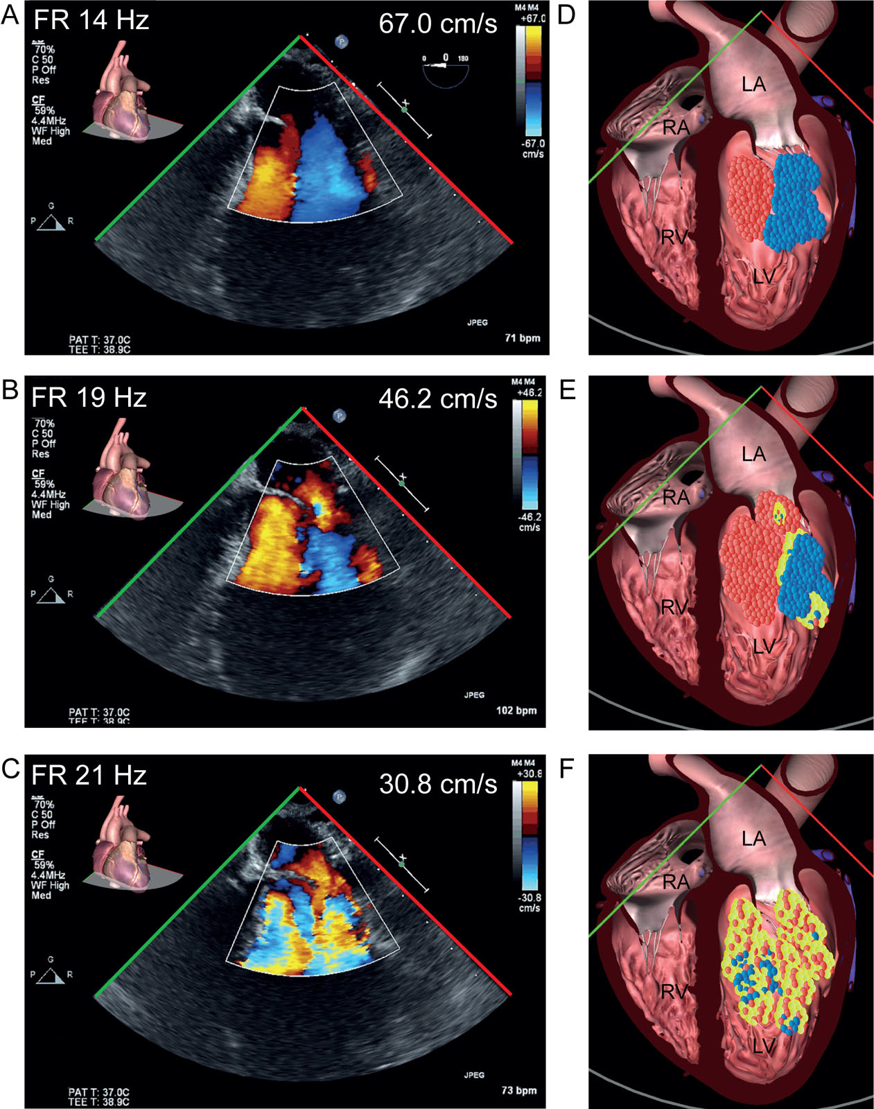
CFI scale and FR. ME 4C views with CFI, in which modifying the color Doppler scale changes the temporal resolution and color display of blood flow. The greater Doppler scale (Nyquist limit) provides less color representation of blood flow with reduced frame rates or PRF. (A) Compare the 2D CFI with Nyquist limits of ±67 cm/s, and reducing the limits to (B) ±46.2 cm/s and (C) ±30.8 cm/s. The temporal resolution and the FR increase by enhancing PRF but exaggerate the color flow pattern of the same blood flows. Abbreviations: 2D, two-dimensional; 4C, four-chamber; CFI, color flow imaging; FR, frame rate; LA, left atrium: LV, left ventricle; ME, mid-esophageal; PRF, pulse repetition frequency; RA, right atrium; RV, right ventricle.
eFigure 3.14CFI scale and FR. ME 4C views with CFI, in which modifying the color Doppler scale changes the temporal resolution and color display of blood flow. The greater Doppler scale (Nyquist limit) provides less color representation of blood flow with reduced frame rates or PRF. (A) Compare the 2D CFI with Nyquist limits of ±67 cm/s, and reducing the limits to (B) ±46.2 cm/s and (C) ±30.8 cm/s. The temporal resolution and the FR increase by enhancing PRF but exaggerate the color flow pattern of the same blood flows. Abbreviations: 2D, two-dimensional; 4C, four-chamber; CFI, color flow imaging; FR, frame rate; LA, left atrium: LV, left ventricle; ME, mid-esophageal; PRF, pulse repetition frequency; RA, right atrium; RV, right ventricle.
eFigure 3.17
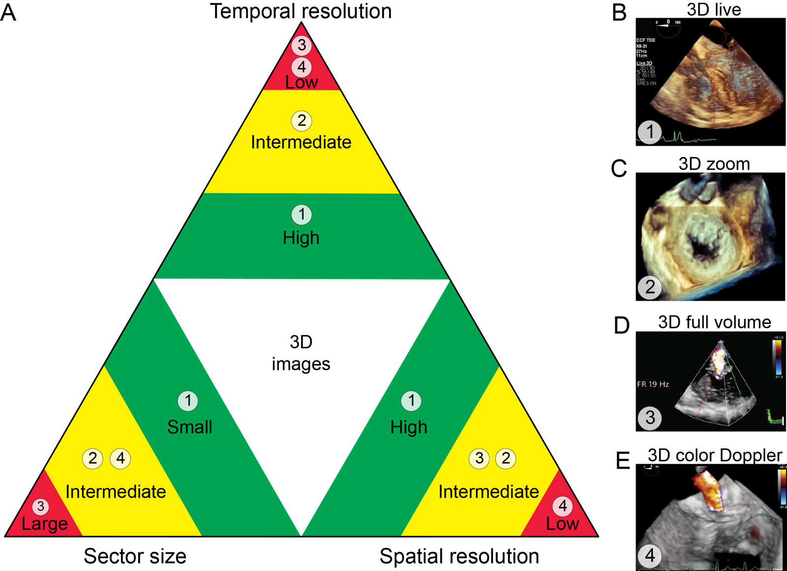
3D imaging factors. (A) 3D image generation implies a balance between temporal resolution, spatial resolution and 3D volume. The spatial resolution depends on the line density. Increased spatial resolution or 3D volume will be at the expense of reducing temporal resolution. (B) 3D live-mode has the lowest sector size, 20-30 Hz temporal resolution and high spatial resolution. (C) 3D Zoom has an intermediate 3D volume, temporal resolution and spatial resolution. (D) 3D full volume has the largest 3D volume, low temporal resolution and intermediate spatial resolution. (E) 3D color flow Imaging has an intermediate 3D volume but because of the superimposed color Doppler the poorest spatial and temporal resolution. Abbreviations: 3D, three-dimension; Hz, hertz; Adapted from Vegas 7 and Mahmood et al. 13
eFigure 3.173D imaging factors. (A) 3D image generation implies a balance between temporal resolution, spatial resolution and 3D volume. The spatial resolution depends on the line density. Increased spatial resolution or 3D volume will be at the expense of reducing temporal resolution. (B) 3D live-mode has the lowest sector size, 20-30 Hz temporal resolution and high spatial resolution. (C) 3D Zoom has an intermediate 3D volume, temporal resolution and spatial resolution. (D) 3D full volume has the largest 3D volume, low temporal resolution and intermediate spatial resolution. (E) 3D color flow Imaging has an intermediate 3D volume but because of the superimposed color Doppler the poorest spatial and temporal resolution. Abbreviations: 3D, three-dimension; Hz, hertz; Adapted from Vegas 7 and Mahmood et al. 13
eFigure 3.19
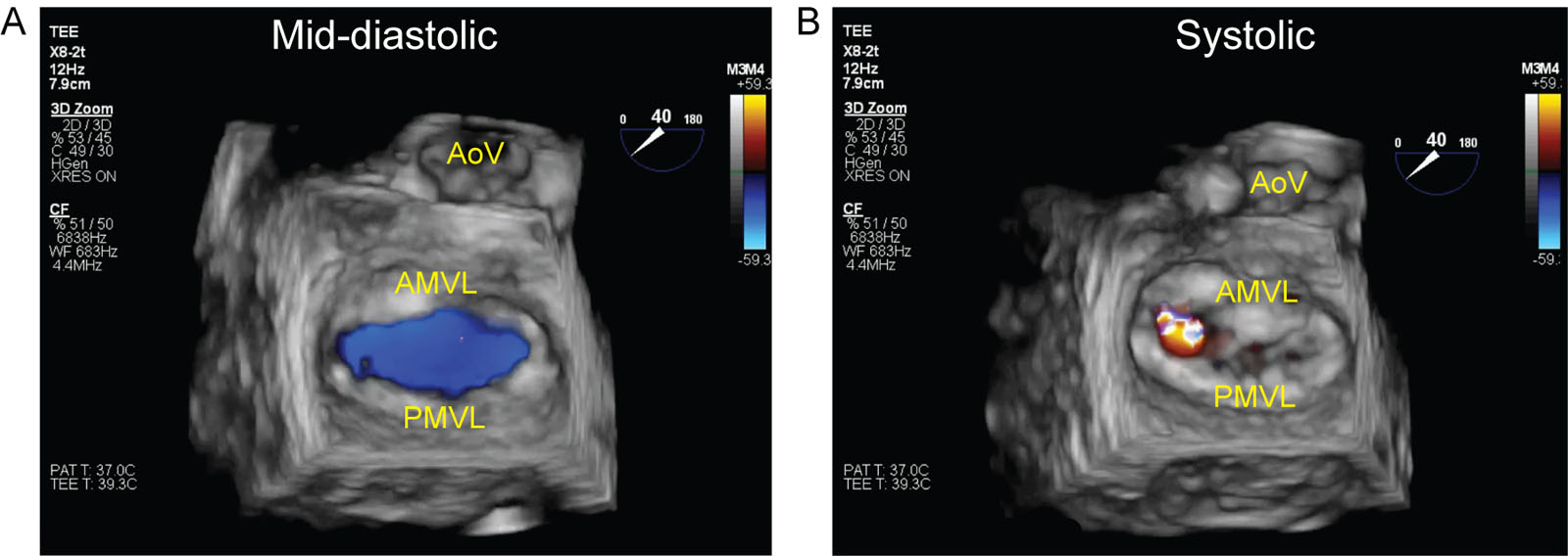
3D CFI with multibeat imaging. (A) 3D CFI mid-diastolic image shows blue-colored transmitral inflow through the opened AMVL and PMVL. (B) 3D CFI mid-systolic image shows a mitral regurgitant jet flow originating from the prolapsed A1 scallop of the AMVL. Abbreviations: 3D, three-dimensional AMVL, anterior mitral valve leaflet; AoV, aortic valve; CFI, color flow imaging; PMVL, posterior mitral valve leaflet.
eFigure 3.193D CFI with multibeat imaging. (A) 3D CFI mid-diastolic image shows blue-colored transmitral inflow through the opened AMVL and PMVL. (B) 3D CFI mid-systolic image shows a mitral regurgitant jet flow originating from the prolapsed A1 scallop of the AMVL. Abbreviations: 3D, three-dimensional AMVL, anterior mitral valve leaflet; AoV, aortic valve; CFI, color flow imaging; PMVL, posterior mitral valve leaflet.
Videos
Chapter 03 Fig01
Chapter 03 Fig9A
Chapter 03 Fig9C
Chapter 03 Fig11AB
Chapter 03 Fig13B
Chapter 03 Fig13C
Chapter 03 Fig13D
Tables
eTable 3.1 Transducer arrays
| Transducer array | Scanning or steering |
Focusing |
Image display |
Mechanical sector |
Motor drive |
Curved lens or element |
Sector |
Linear array |
Electronic sequencing |
Electronic phasing |
Rectangular |
Phased linear array |
Electronic sequencing |
Electronic phasing |
Parallelogram |
Convex array |
Electronic sequencing |
Intrinsic property of |
Sector |
Phased convex array |
Electronic sequencing |
Electronic phasing |
Sector |
Annular array |
Motor drive |
Electronic phasing |
Sector |
Vector array |
Electronic sequencing |
Electronic phasing |
Sector |
eTable 3.2 Axial resolution, penetration, and frequency
| Frequency (MHz) | Depth (cm) |
Axial resolution (mm) |
2.0 |
30 |
0.77 |
3.5 |
17 |
0.44 |
5.0 |
12 |
0.31 |
7.5 |
8 |
0.20 |
10.0 |
6 |
0.15 |
15.0 |
4 |
0.10 |
eTable 3.3 Operating frequencies of different ultrasound transducers
| Transducer type | Probe frequency (MHz) |
Transthoracic probe |
|
|
7.5–12.0 |
|
5.0–7.5 |
|
2.5–3.5 |
Transesophageal probe |
3.0–7.0 |
Intravascular ultrasound |
10.0–40.0 |