Figures
eFigure 4.3
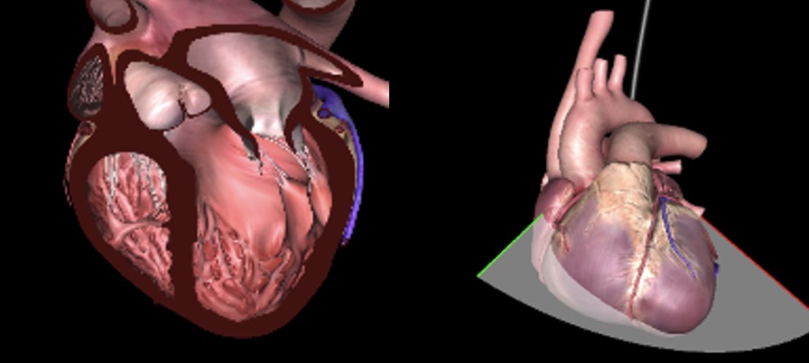
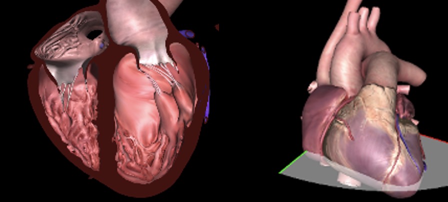
Triplane Multiplane TEE imaging. (A) Simultaneous triplane imaging with the primary imaging plane as the ME 4C view at 4°, and the secondary imaging planes as the ME MC view at 64° and the ME LAX view at 124°. Abbreviations: 4C, four-chamber; LAX, long-axis; ME, mid-esophageal, MC, mitral commissural; TEE, transesophageal echocardiography.
eFigure 4.3Triplane Multiplane TEE imaging. (A) Simultaneous triplane imaging with the primary imaging plane as the ME 4C view at 4°, and the secondary imaging planes as the ME MC view at 64° and the ME LAX view at 124°. Abbreviations: 4C, four-chamber; LAX, long-axis; ME, mid-esophageal, MC, mitral commissural; TEE, transesophageal echocardiography.
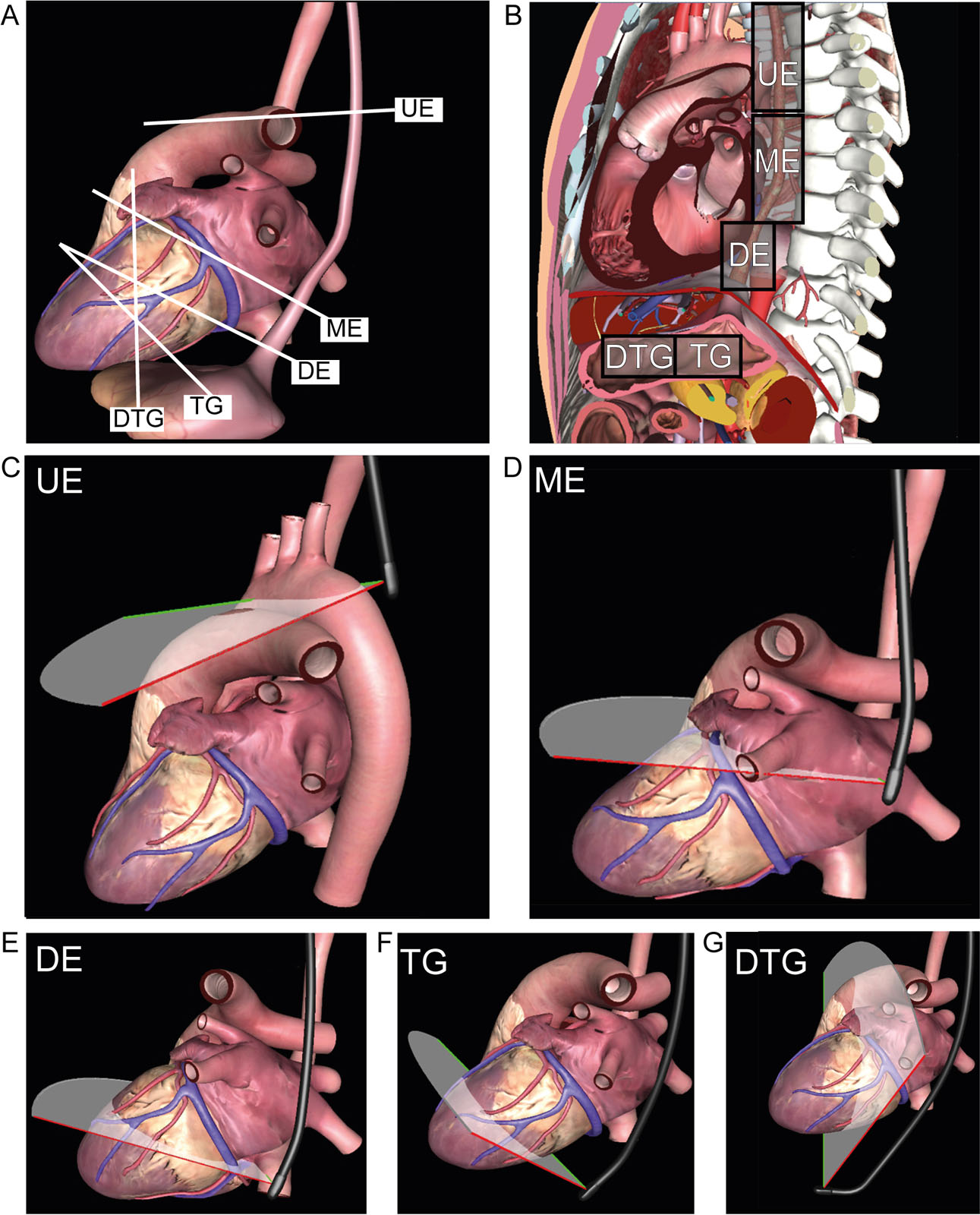
eFigure 4.39
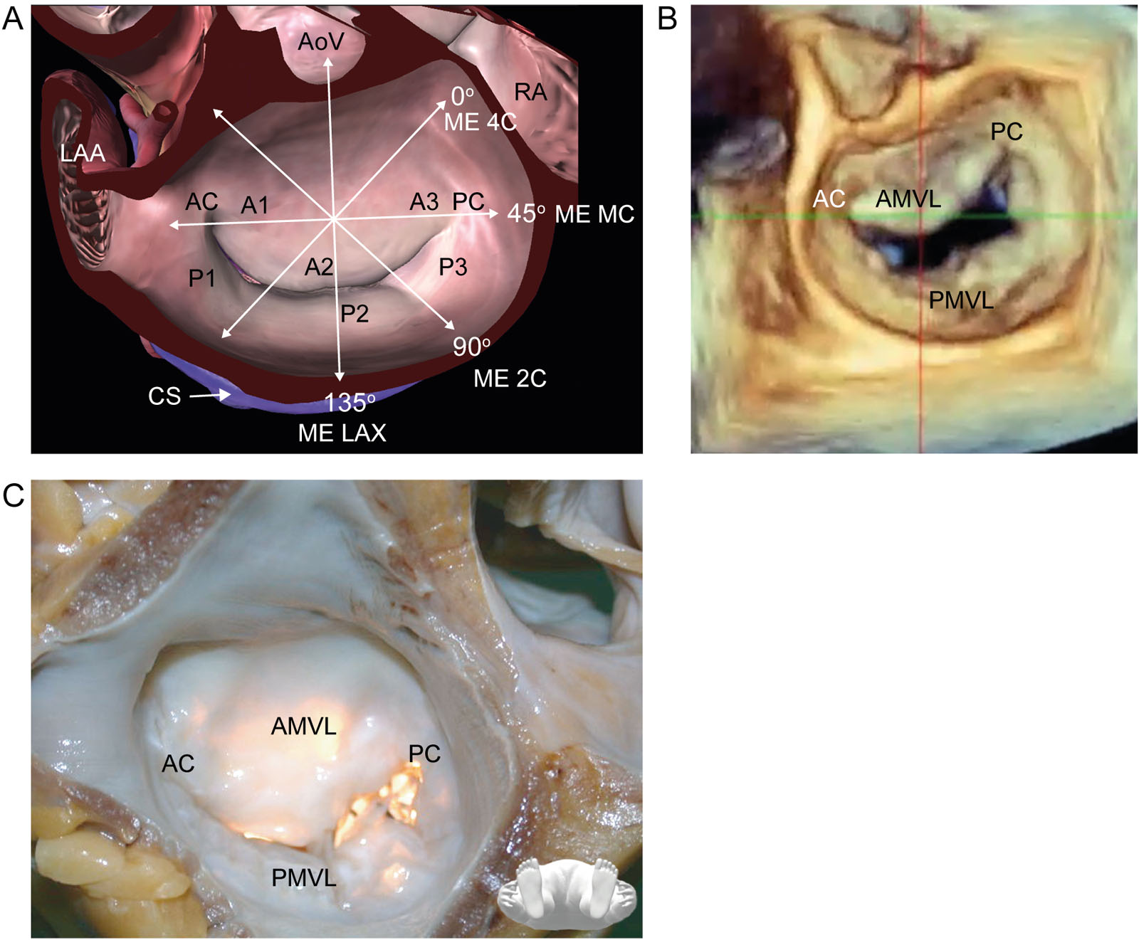
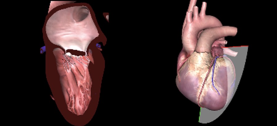
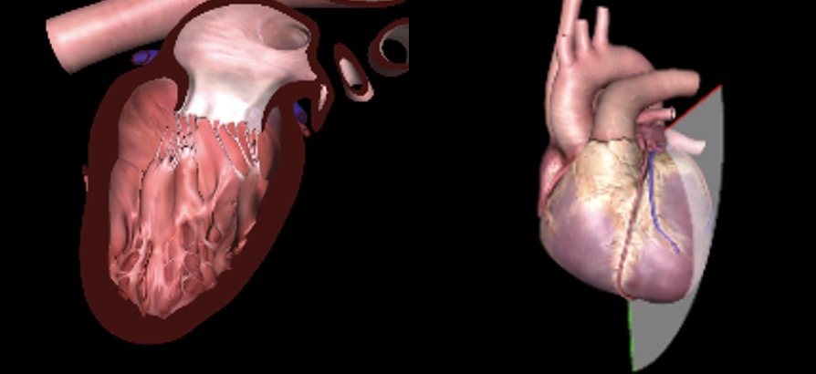
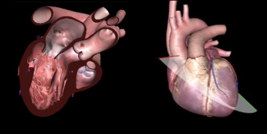
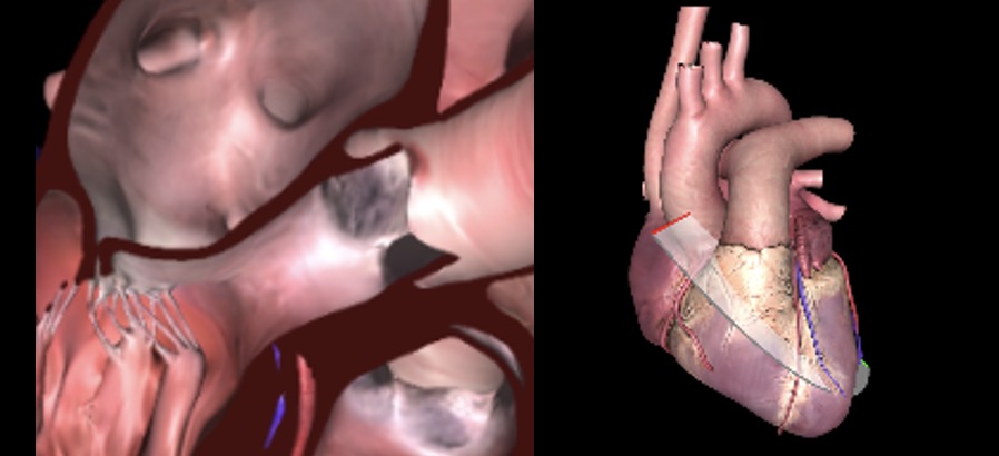
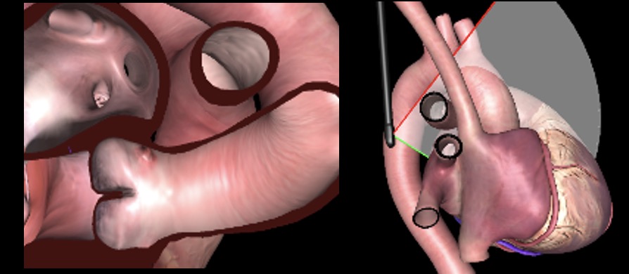
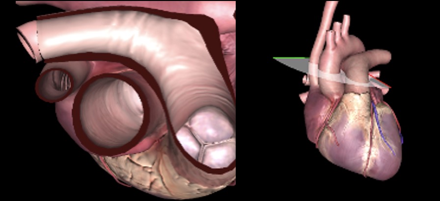
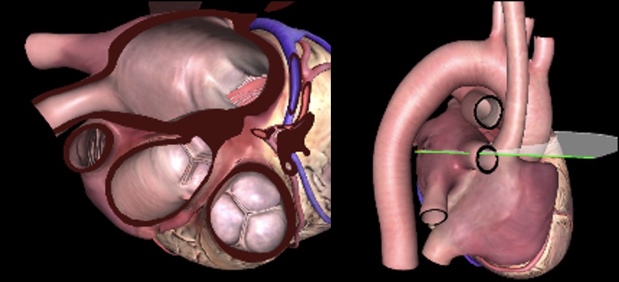
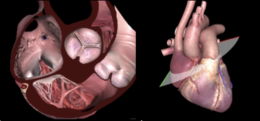
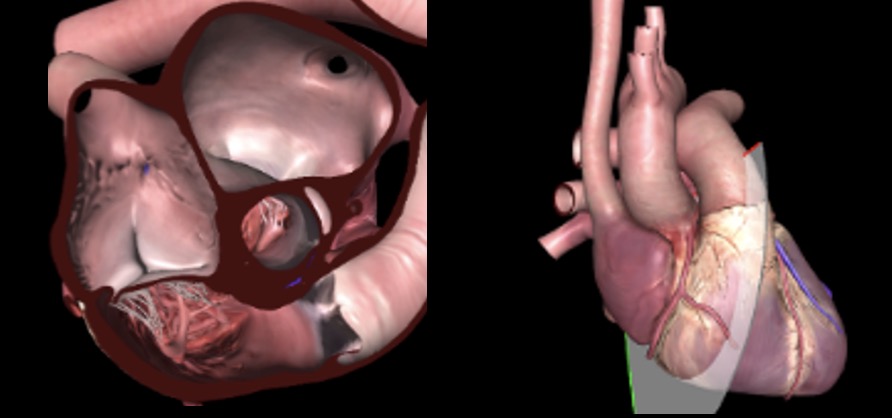
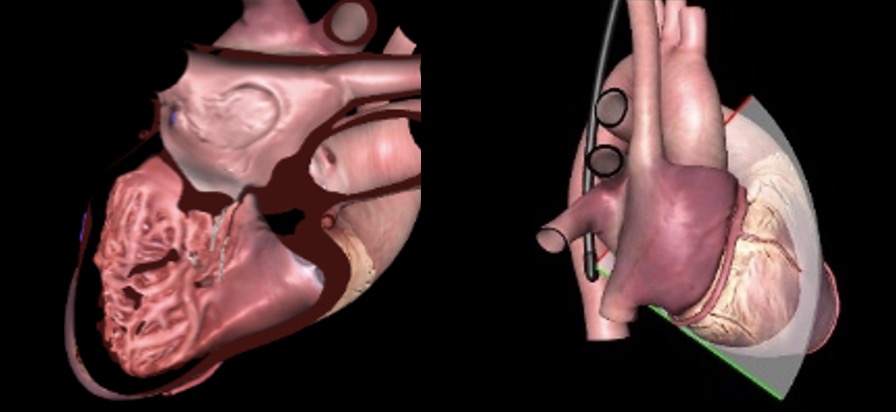
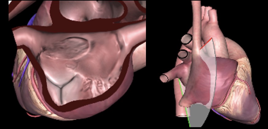
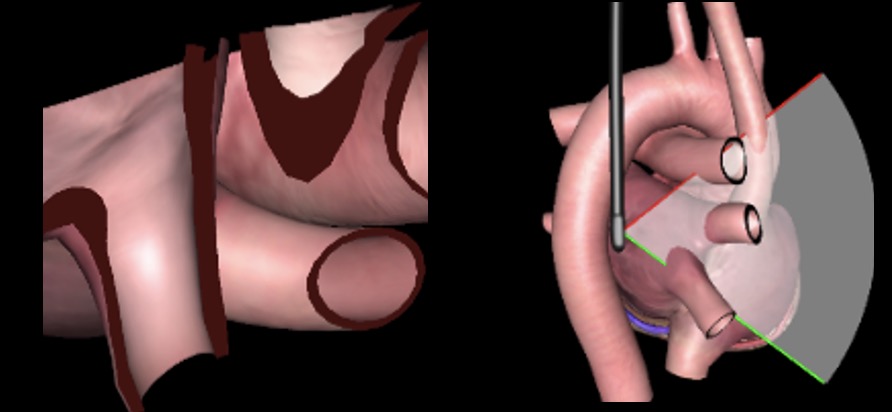
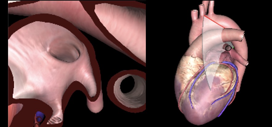
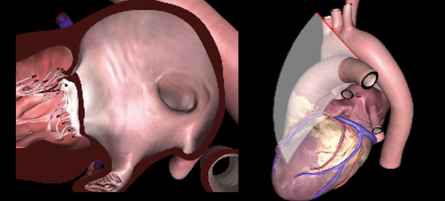
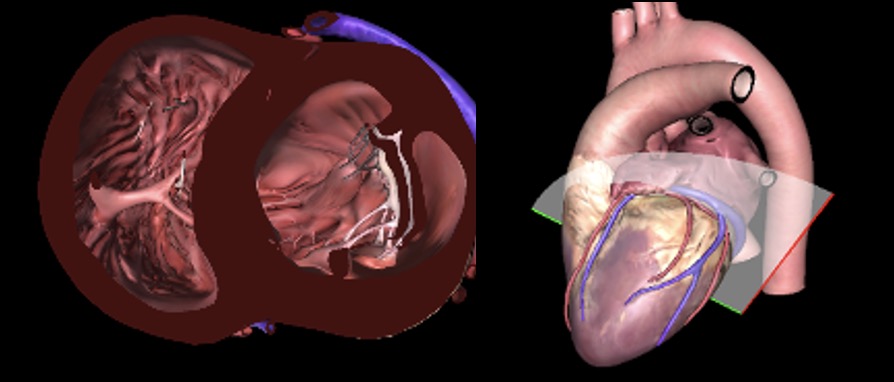
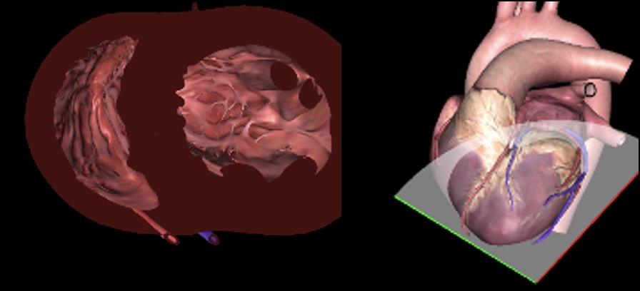
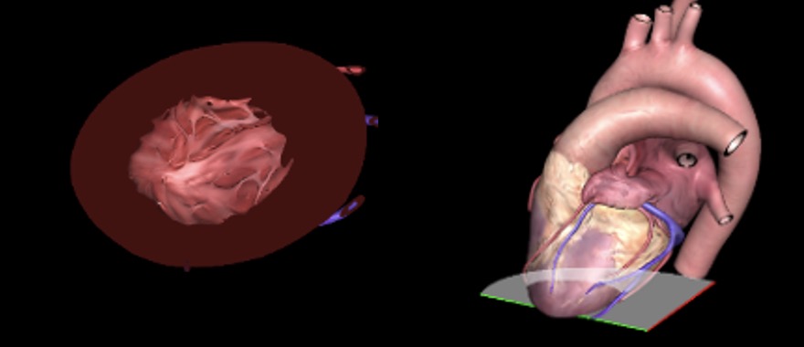
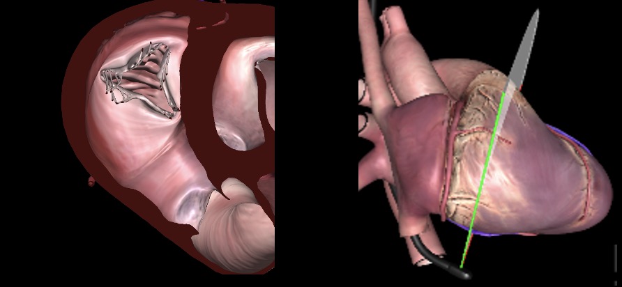
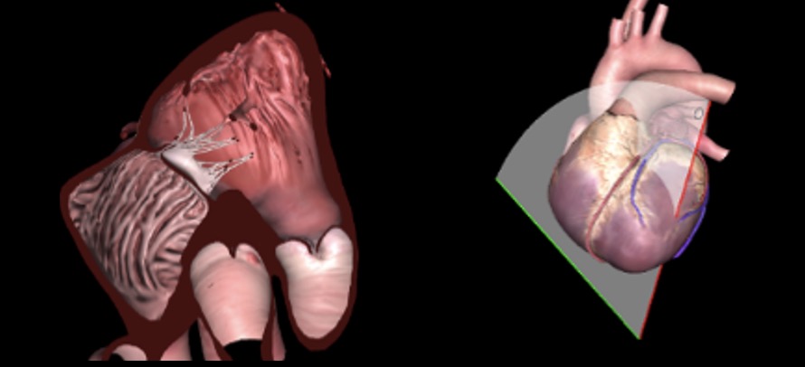
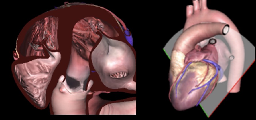
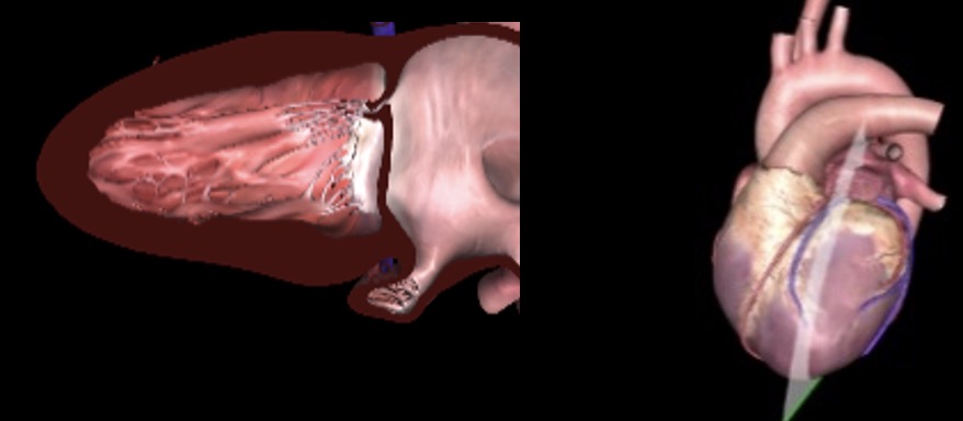
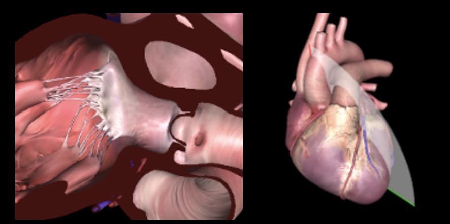
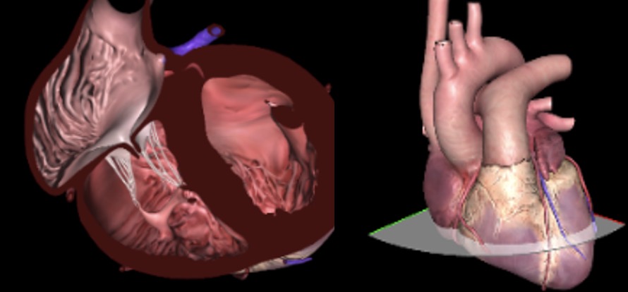
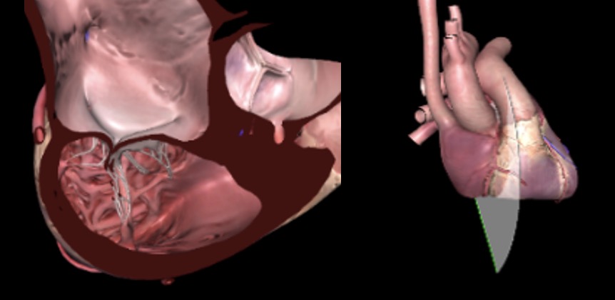
Mitral valve. (A) This is a schematic diagram of the MV with labeled leaflet scallops and segments. (B, C) Corresponding 3D en-face and anatomic images of the MV. The region of the MV leaflets that appear in a 2D image varies with the relation of the heart to the esophagus. Abbreviations: 2C, two-Chambers; 2D, two-dimensional; 3D, three-dimensional; A, anterior MV segments; AC, anterior commissure; AMVL, anterior MV leaflet; AoV, aortic valve; CS, coronary sinus; LAA, left atrial appendage; LAX, long-axis; MC, mitral commissural; ME, mid-esophageal; P, posterior MV segments; PC, posterior commissure; PMVL, posterior MV leaflet; RA, right atrium.
eFigure 4.39Mitral valve. (A) This is a schematic diagram of the MV with labeled leaflet scallops and segments. (B, C) Corresponding 3D en-face and anatomic images of the MV. The region of the MV leaflets that appear in a 2D image varies with the relation of the heart to the esophagus. Abbreviations: 2C, two-Chambers; 2D, two-dimensional; 3D, three-dimensional; A, anterior MV segments; AC, anterior commissure; AMVL, anterior MV leaflet; AoV, aortic valve; CS, coronary sinus; LAA, left atrial appendage; LAX, long-axis; MC, mitral commissural; ME, mid-esophageal; P, posterior MV segments; PC, posterior commissure; PMVL, posterior MV leaflet; RA, right atrium.
Videos
Chapter 04 Fig01
Chapter 04 Fig02A
Chapter 04 Fig02C
Chapter 04 Fig03
Chapter 04 Fig05A
Chapter 04 Fig05C
Chapter 04 Fig05E
Chapter 04 Fig06A
Chapter 04 Fig06B
Chapter 04 Fig06C
Chapter 04 Fig06E
Chapter 04 Fig07A
Chapter 04 Fig07B
Chapter 04 Fig07C
Chapter 04 Fig07E
Chapter 04 Fig07F
Chapter 04 Fig08A
Chapter 04 Fig08C
Chapter 04 Fig08E
Chapter 04 Fig09A
Chapter 04 Fig09B
Chapter 04 Fig09C
Chapter 04 Fig09E
Chapter 04 Fig09F
Chapter 04 Fig10A
Chapter 04 Fig10B
Chapter 04 Fig10C
Chapter 04 Fig10E
Chapter 04 Fig11A
Chapter 04 Fig11B
Chapter 04 Fig11C
Chapter 04 Fig11E
Chapter 04 Fig12A
Chapter 04 Fig12B
Chapter 04 Fig12C
Chapter 04 Fig12E
Chapter 04 Fig12F
Chapter 04 Fig13A
Chapter 04 Fig13B
Chapter 04 Fig13C
Chapter 04 Fig13E
Chapter 04 Fig14A
Chapter 04 Fig14B
Chapter 04 Fig14C
Chapter 04 Fig14E
Chapter 04 Fig14F
Chapter 04 Fig15A
Chapter 04 Fig15B
Chapter 04 Fig15C
Chapter 04 Fig15E
Chapter 04 Fig15F
Chapter 04 Fig16A
Chapter 04 Fig16B
Chapter 04 Fig16C
Chapter 04 Fig16E
Chapter 04 Fig17A
Chapter 04 Fig17B
Chapter 04 Fig17C
Chapter 04 Fig17E
Chapter 04 Fig18A
Chapter 04 Fig18B
Chapter 04 Fig18C
Chapter 04 Fig18E
Chapter 04 Fig18F
Chapter 04 Fig19A
Chapter 04 Fig19B
Chapter 04 Fig19C
Chapter 04 Fig19E
Chapter 04 Fig20A
Chapter 04 Fig20B
Chapter 04 Fig20C
Chapter 04 Fig20E
Chapter 04 Fig21A
Chapter 04 Fig21B
Chapter 04 Fig21C
Chapter 04 Fig21E
Chapter 04 Fig22A
Chapter 04 Fig22B
Chapter 04 Fig22C
Chapter 04 Fig22E
Chapter 04 Fig22F
Chapter 04 Fig23A
Chapter 04 Fig23B
Chapter 04 Fig23C
Chapter 04 Fig23E
Chapter 04 Fig23F
Chapter 04 Fig24A
Chapter 04 Fig24B
Chapter 04 Fig24C
Chapter 04 Fig24E
Chapter 04 Fig25A
Chapter 04 Fig25B
Chapter 04 Fig25C
Chapter 04 Fig25D
Chapter 04 Fig25E
Chapter 04 Fig25F
Chapter 04 Fig26A
Chapter 04 Fig26B
Chapter 04 Fig26C
Chapter 04 Fig26D
Chapter 04 Fig26E
Chapter 04 Fig26F
Chapter 04 Fig27A
Chapter 04 Fig27B
Chapter 04 Fig27C
Chapter 04 Fig27E
Chapter 04 Fig27F
Chapter 04 Fig28A
Chapter 04 Fig28B
Chapter 04 Fig28C
Chapter 04 Fig28E
Chapter 04 Fig29A
Chapter 04 Fig29B
Chapter 04 Fig29C
Chapter 04 Fig29E
Chapter 04 Fig29F
Chapter 04 Fig30A
Chapter 04 Fig30B
Chapter 04 Fig30C
Chapter 04 Fig30E
Chapter 04 Fig30F
Chapter 04 Fig31A
Chapter 04 Fig31B
Chapter 04 Fig31C
Chapter 04 Fig31E
Chapter 04 Fig31F
Chapter 04 Fig32A
Chapter 04 Fig32B
Chapter 04 Fig32C
Chapter 04 Fig32Eok
Chapter 04 Fig32F
Chapter 04 Fig33A
Chapter 04 Fig33B
Chapter 04 Fig33C
Chapter 04 Fig33E
Chapter 04 Fig34A
Chapter 04 Fig34B
Chapter 04 Fig34C
Chapter 04 Fig34E
Chapter 04 Fig34F
Chapter 04 Fig35B
Chapter 04 Fig36A
Chapter 04 Fig36B
Chapter 04 Fig36C
Chapter 04 Fig36F
Chapter 04 Fig37A
Chapter 04 Fig37B
Chapter 04 Fig37C
Chapter 04 Fig37E
Chapter 04 Fig37F
Chapter 04 Fig38A
Chapter 04 Fig38B
Chapter 04 Fig38C
Chapter 04 Fig38D
Chapter 04 Fig38E
Chapter 04 Fig38F
Chapter 04 Fig39A
Chapter 04 Fig39B
Tables
eTable 4.1 TEE probe orientation
| Orientation (± 15-30°) |
Description and features of view |
0° |
Transverse plane to probe shaft long axis, and horizontal to plane of the body |
45° |
SAX view of cardiac basal structures (e.g., aortic valve) |
90° |
Longitudinal plane parallel to probe shaft LAX and sagittal (vertical) to plane of the body |
135° |
LAX view of cardiac structures (LV and LVOT) |
180° |
Mirror image of the 0° transverse plane |
Abbreviations: LAX, long axis; LV, left ventricle; LVOT, left ventricular outflow tract; SAX, short axis. |
|
eTable 4.2 TEE views
|
TEE View | Icon |
View-Technique |
Utility |
||
1 |
|
ME 5C |
Chambers: LA, LV, LVOT, RA, RV |
||
2 |
|
ME 4C |
Chambers: LA, LV, RA, RV |
||
3 |
|
ME Mitral commissural |
Chambers: LA, LV |
||
4 |
|
ME 2C |
Chambers: LA, LV |
||
5 |
|
ME LAX |
Chambers: LA, LV, LVOT, RVOT |
||
6 |
|
ME AoV LAX |
Chambers: LA, LVOT, RVOT |
||
7 |
|
ME Ascending aorta LAX |
Vessels: Asc Ao, RPA |
||
8 |
|
ME Ascending Aorta SAX |
Vessels: Asc Ao, PA, SVC |
||
9 |
|
ME RUPV |
Vessels: Asc Ao, RUPV, SVC |
||
10 |
|
ME AoV SAX |
Chambers: LA, RA, RV, RVOT |
||
11 |
|
ME RV Inflow-Outflow |
Chambers: LA, RA, RV, RVOT |
||
12 |
|
ME Modified Bicaval TV |
Chambers: LA, RA, RAA, RV |
||
13 |
|
ME Bicaval |
Chambers: LA, RA, RAA |
||
14a |
|
ME RUPV |
Chamber: LA |
||
14b |
|
ME LUPV |
Chamber: LA, LAA |
||
15 |
|
ME LAA |
Chamber: LAA |
||
16 |
|
TG Basal SAX |
Chambers: LV, RV |
||
17 |
|
TG Mid papillary SAX |
Chambers: LV, RV |
||
18 |
|
TG Apical SAX |
Chambers: LV, RV |
||
19 |
|
TG RV basal SAX |
Chambers: LV, RV, RVOT |
||
20 |
|
TG RV Inflow outflow |
Chambers: RA, RV, RVOT |
||
21 |
|
DTG 5C |
Chambers: LA, LV, LVOT, RA, RV |
||
22 |
|
TG 2C LAX |
Chambers: LA, LV, LAA |
||
23 |
|
TG RV inflow |
Chambers: RA, RV, RVOT |
||
24 |
|
TG LAX |
Chambers: LA, LV, LVOT |
||
25 |
|
Descending aorta SAX |
Vessels: Des Ao, hemiazygos and azygos vein, intercostal arteries |
||
26 |
|
Descending aorta LAX Angle: 90-100° |
Vessels: Des Ao, intercostal arteries |
||
27 |
|
UE Aortic arch LAX |
Vessels: Aortic arch, innominate and hemiazygos vein |
||
28 |
|
UE Aortic arch SAX |
Valve: PV |
||
29 |
|
DE 2C RV |
Chambers: RA, RV |
||
30 |
|
DE RV inflow-outflow |
Chambers: RA, RV, RVOT |
||
Additional cardiac and extra-cardiac views (TELUS and TEGAUS) appear in other chapters. |
|||||
Abbreviations: 2C, two-chamber; 4C, four-chamber; 5C, five-chamber; Asc, ascending; Ao, aorta; AoV, aortic valve; CO, cardiac output; DE, deep-esophageal; Des, descending; DTG, deep transgastric; IAS, interatrial septum; IVC, inferior vena cava; IVS, interventricular septum; LA, left atrium; LAA, left atrial appendage; LAX, long-axis; LMCA, left main coronary artery; LV, left ventricle; LVOT, left ventricular outflow tract; ME, mid-esophageal; MV, mitral valve; PA, pulmonary artery; PV, pulmonic valve; RA, right atrium; RAA, right atrial appendage; RPA, right pulmonary artery; RV, right ventricle; RVOT, right ventricular outflow tract; RVOT, right ventricular outflow tract; RVSP, right ventricular systolic pressure; SAX, short-axis; SVC, superior vena cava; SPAP, systolic pulmonary artery pressure; TELUS, transesophageal lung ultrasound; TG, transgastric; TGAUS, transgastric abdominal ultrasound; TR, tricuspid regurgitation; TV, tricuspid valve; UE, upper esophageal; VTI, velocity time integral. Adapted from Hahn et al. |
|||||
eTable 4.3 Normal filling pressures in cardiac chambers and great vessels
| Chamber | Pressure (mmHg) |
||
Systolic |
Diastolic |
Mean |
|
RA |
1–8 |
||
RV |
15–30 |
1–8 |
|
PA |
15–30 |
4–12 |
9–18 |
LA |
2–12 |
||
LV |
100–140 |
3–12 |
|
Aorta |
100–140 |
60–90 |
70–105 |
Abbreviations: LA, left atrium; LV, left ventricle; PA, pulmonary artery; RA, right atrium; RV, right ventricle. |
|||