Figures
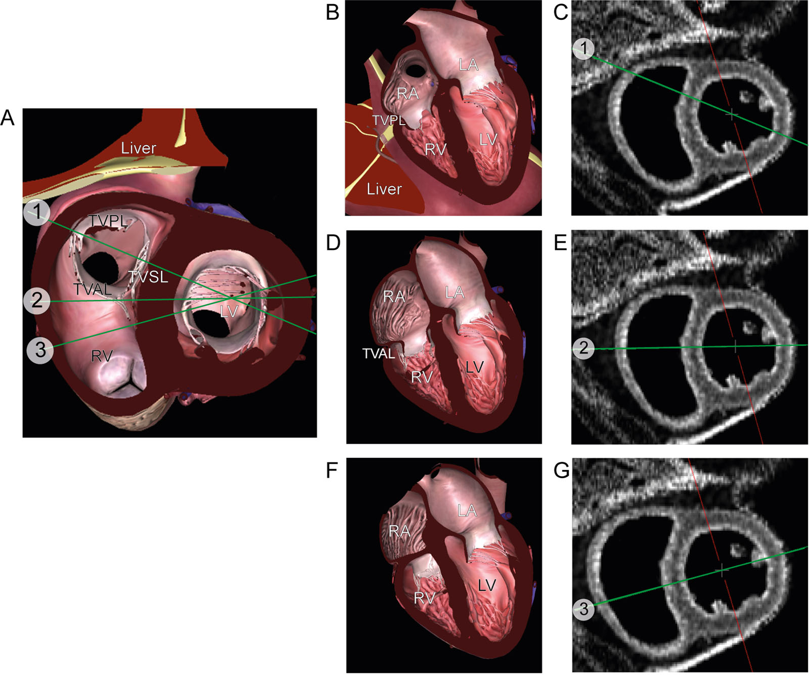
eFigure 5.4
Pitfall in RV basal diameter measurement. (A) TG basal SAX view showing ME RV 4C views at 3 different positions: (B, C) inflow or position#1 (D, E) middle or position #2 and (F, G) and RV outflow tract or position #3. Although there is a similarity in those positions, the RVD will vary depending on its location in the basal RV. The largest RVD at the level of the TVAL (position #2)) should be selected for measurement. Abbreviations: 4C, four-chamber; LA, left atrium; LV, left ventricle; ME, mid-esophageal; RA, right atrium; RV, right ventricular; RVD, right ventricular diameter; SAX, short-axis; TG, transgastric; TVAL, tricuspid valve anterior leaflet; TVPL, tricuspid valve posterior leaflet; TVSL, tricuspid valve septal leaflet. Adapted from Rudski 41 and Lang et al.6
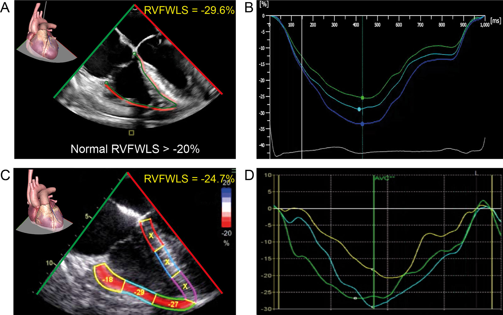
eFigure 5.18
RV systolic strain. ME 4C analyzed with speckle tracking echocardiography shows the RVFWLS averaging the three segments of the free wall using (A, B) Philips and (C, D) GE software. Abbreviations: 4C, four-chambers; ME, mid-esophageal; RV, right ventricular; RVFWLS, right ventricular free wall longitudinal strain.