Figures
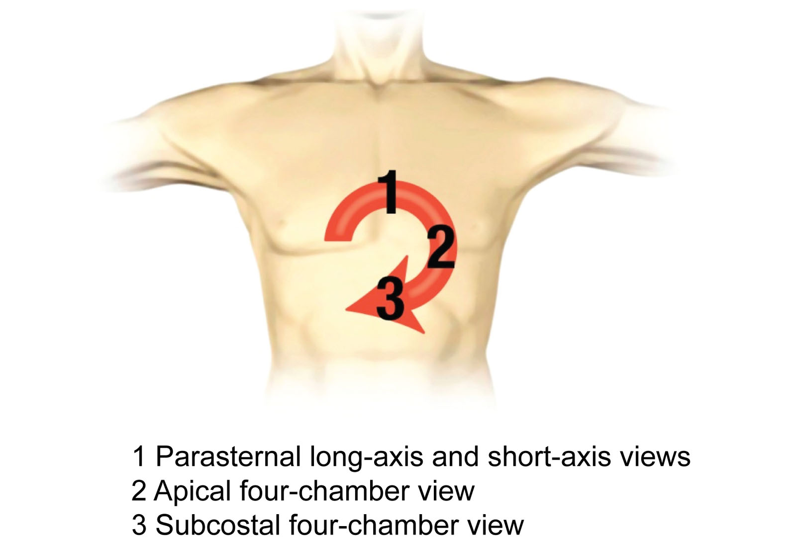
eFigure 6.1
Focused cardiac ultrasound study (FOCUS). The FOCUS exam includes the most important echocardiographic views with scanning in a systematic, clockwise fashion, from three main areas: the parasternal (1), apical (2), and subcostal (3) areas. Adapted from the FOCUS pocket guide, with permission from ICCU Imaging Inc.
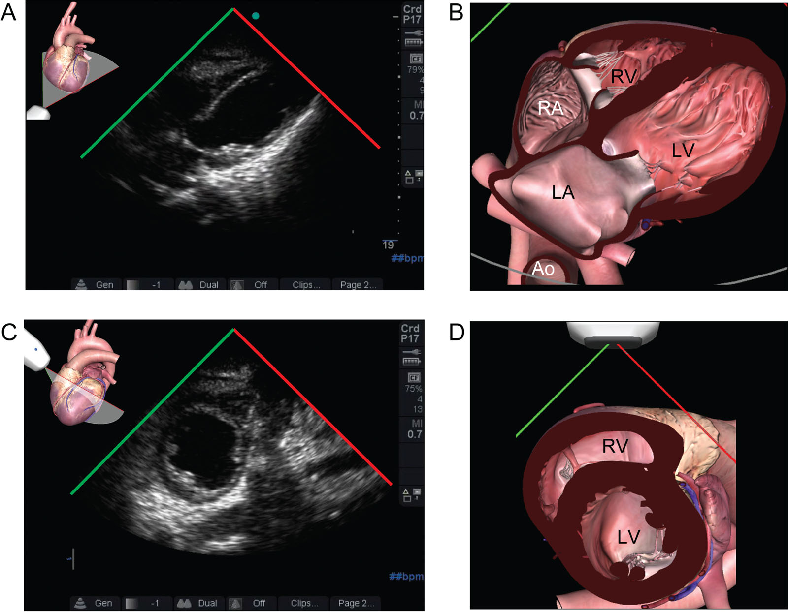
eFigure 6.5
Takotsubo syndrome. Acute hemodynamic instability occurs in a 73-year-old woman after duodenal perforation. (A, B) A focused cardiac ultrasound study (FOCUS) exam from a subcostal view revealed severe left ventricular (LV) dysfunction with basal sparing typical of Takotsubo syndrome. (C, D) A parasternal SAX view confirms the abnormal LV function. Abbreviations: LA, left atrium; RA, right atrium; RV, right ventricle; SAX, short-axis.
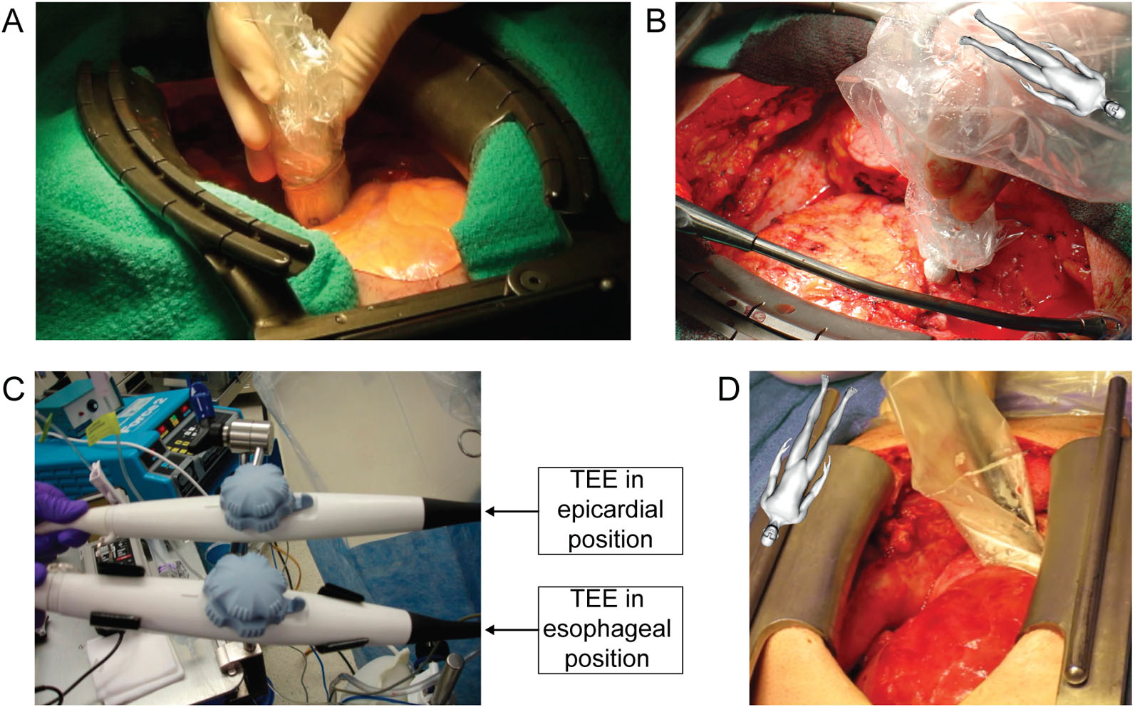
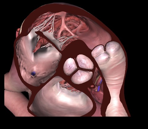
eFigure 6.10
Methods used for epicardial echocardiography. (A, B) A trained cardiac surgeon can manipulate high-frequency transthoracic and linear probes in sterile sheaths on the epicardium to identify the key structures. (C, D) In certain cases, an experienced echocardiographer can use a TEE probe in a sterile sheath that the surgeon positions on the epicardium to enable epicardial echocardiography. Abbreviation: TEE, transesophageal echocardiography. Source: Courtesy of Dr. Michel Pellerin.
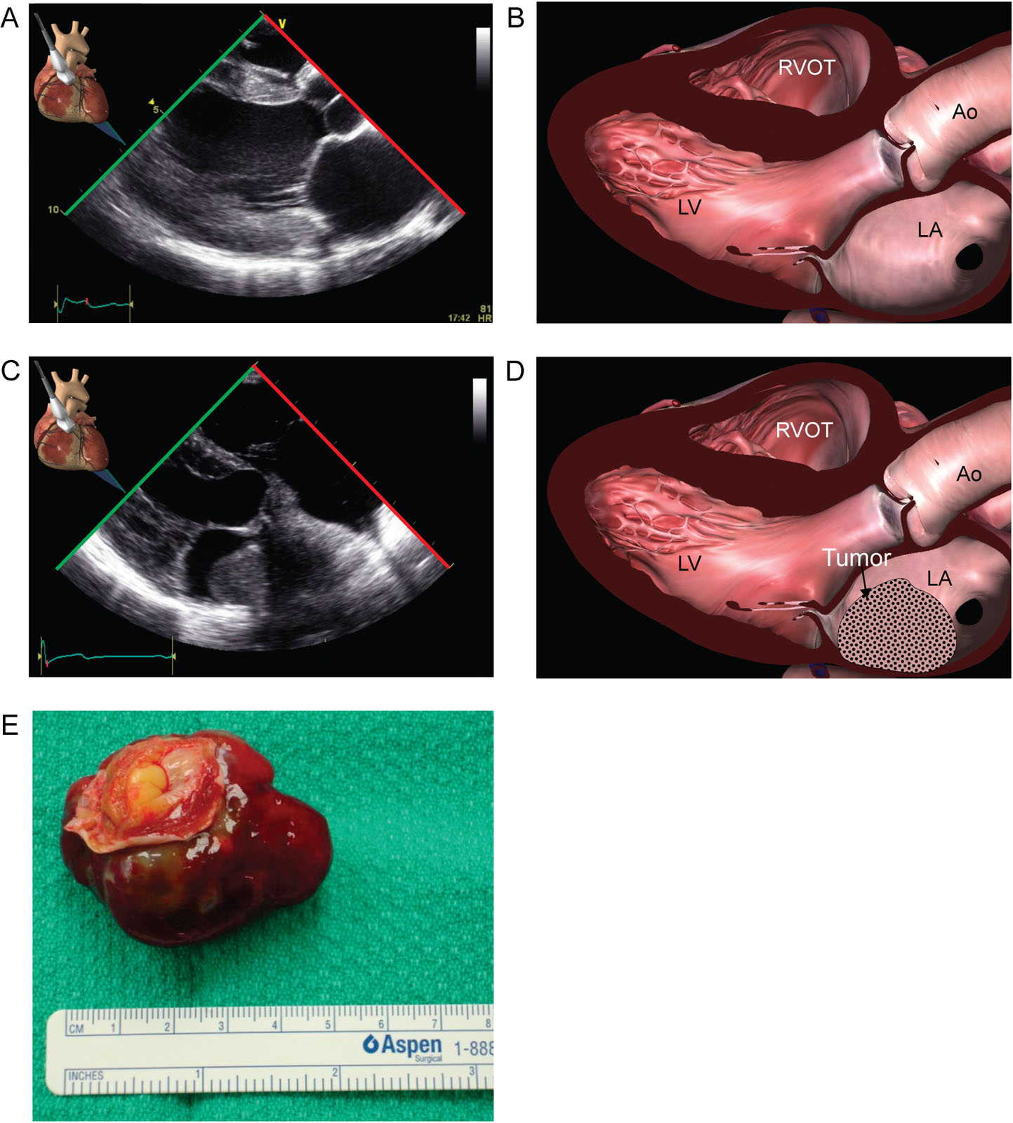
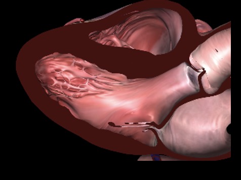
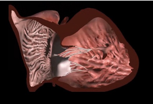
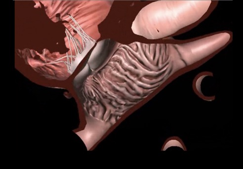
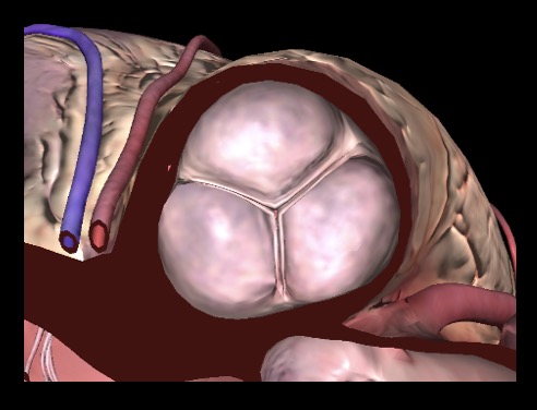
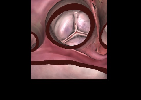
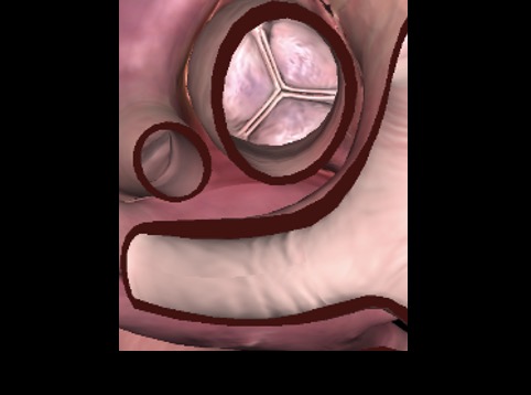
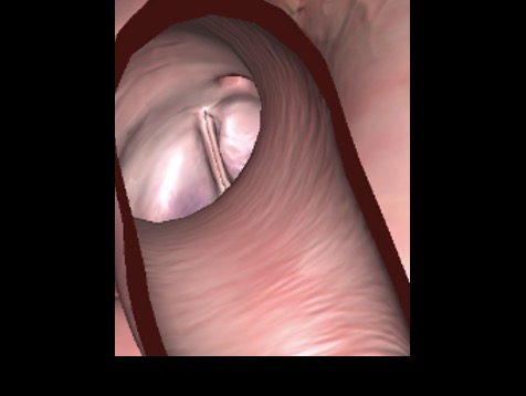
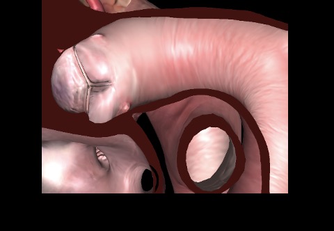
Figure 6.25
Epicardial LV LAX view#5. (A, B) This view is equivalent to the transthoracic parasternal LV LAX view. Probe placement is on the anterior RV with a marker towards the right shoulder. From the epicardial LV mid-SAX view, angling the ultrasound beam superiorly and rotating the probe toward the patient’s right shoulder generates the epicardial LV LAX view. This view evaluates the inferolateral and anteroseptal walls of the LV and the RV, LA, LVOT, aortic valve, and mitral valve. Color flow Doppler can assess for aortic regurgitation, mitral regurgitation, ventricular septal defects, LVOT obstruction, or systolic anterior motion of the mitral valve using this view. (C, D) An epicardial LV LAX view before myxoma removal localizes fixation of the tumor on the interatrial septum. (E) Pathological specimen is shown. Abbreviations: Ao, aorta; LA, left atrium; LAX, long-axis; LV, left ventricle; LVOT, left ventricular outflow tract; RVOT, right ventricular outflow tract. Photo E courtesy of Dr. Michel Pellerin. Adapted from Reeves et al. 73.
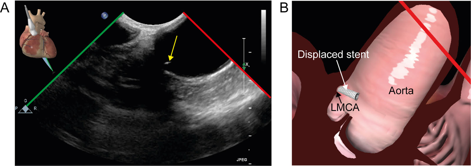
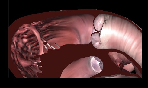
eFigure 6.29
Broken stent in LMCA (A, B) Epicardial view of a broken stent in the LMCA and the epiaortic view shows a small linear hyper-echoic artefact close to the LMCA orifice. Abbreviations: LMCA, left main coronary artery.
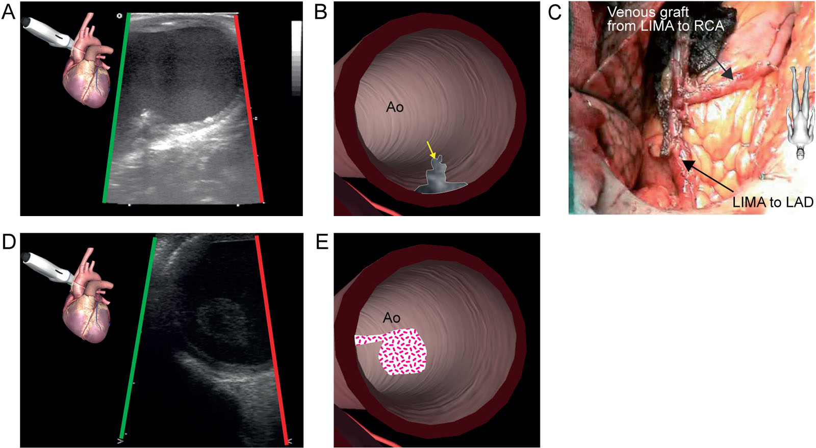
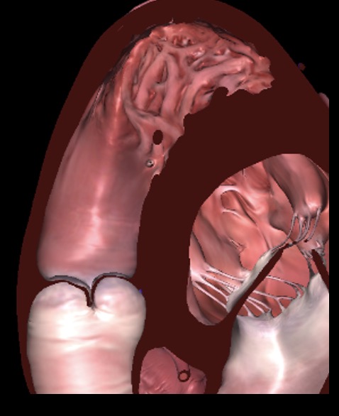
Figure 6.39
Mobile atheroma, (A, B) Epiaortic images of the aortic arch in zone 2 reveal a mobile atheroma (Grade 5) in a 74-year-old woman before cardiopulmonary bypass. (C) To avoid aortic clamping during off-pump coronary artery bypass grafting, the surgeon anastomosed the saphenous vein graft between the RCA and the LIMA graft rather than onto the aorta, as is usually the case. (D,E) Unexpected floating plaque or thrombus in the ascending aorta. Abbreviations: Ao, aorta; LAD, left anterior descending coronary artery; LIMA, left internal mammary artery; RCA, right coronary artery. Source: Photo C courtesy of Dr. Louis P. Perrault.
Videos
Chapter 06 Fig02A
Chapter 06 Fig02B
Chapter 06 Fig02C
Chapter 06 Fig02D
Chapter 06 Fig02E
Chapter 06 Fig02F
Chapter 06 Fig02G
Chapter 06 Fig02H
Chapter 06 Fig03B
Chapter 06 Fig03C
Chapter 06 Fig03D
Chapter 06 Fig04A
Chapter 06 Fig04D
Chapter 06 Fig05A
Chapter 06 Fig05C
Chapter 06 Fig06A
Chapter 06 Fig07A
Chapter 06 Fig08A
Chapter 06 Fig08B
Chapter 06 Fig09A
Chapter 06 Fig09B
Chapter 06 Fig10D
Chapter 06 Fig11A
Chapter 06 Fig12A
Chapter 06 Fig12C
Chapter 06 Fig13A
Chapter 06 Fig13B
Chapter 06 Fig13DE
Chapter 06 Fig14A
Chapter 06 Fig15A
Chapter 06 Fig16A
Chapter 06 Fig17A
Chapter 06 Fig18A
Chapter 06 Fig19A
Chapter 06 Fig20A
Chapter 06 Fig21A
Chapter 06 Fig22A
Chapter 06 Fig22B
Chapter 06 Fig23A
Chapter 06 Fig23C
Chapter 06 Fig24A
Chapter 06 Fig24C
Chapter 06 Fig25A
Chapter 06 Fig25C
Chapter 06 Fig25C1
Chapter 06 Fig26A
Chapter 06 Fig27A
Chapter 06 Fig28A
Chapter 06 Fig28C
Chapter 06 Fig28D
Chapter 06 Fig29A
Chapter 06 Fig31B
Chapter 06 Fig31C
Chapter 06 Fig31D
Chapter 06 Fig31E
Chapter 06 Fig32B
Chapter 06 Fig33A
Chapter 06 Fig34BC
Chapter 06 Fig35B
Chapter 06 Fig36B
Chapter 06 Fig37BC
Chapter 06 Fig38BC
Chapter 06 Fig39A
Chapter 06 Fig39C
Chapter 06 Fig39D
Tables
x Epicardial and epiaortic views
| Epicardial Views | View-Technique | Utility/Disadvantages |
|
||||||||
1 |
 |
Epicardial AoV SAX |
|
|
|||||||
2 |
 |
Epicardial AoV LAX |
|
|
|||||||
3 |
 |
Epicardial LV basal SAX |
|
|
|||||||
4 |
 |
Epicardial LV mid SAX |
|
|
|||||||
5 |
 |
Epicardial LV LAX |
|
|
|||||||
6 |
 |
Epicardial LV 2C |
|
|
|||||||
7 |
 |
Epicardial RVOT |
|
|
|||||||
Alternative epicardial views |
|
||||||||||
Alt#1 |
 |
Epicardial alternative RV inflow |
|
|
|||||||
Alt#2 |
 |
Epicardial alternative RA and bicaval views Orientation marker points cephalad |
|
|
|||||||
Alt#3 |
 |
Epicardial alternative TV SAX |
|
|
|||||||
Alt#4 |
 |
Epicardial alternative PV SAX |
|
|
|||||||
Alt#5 |
 |
Epicardial alternative PV LAX |
|
|
|||||||
Alt#6 |
 |
Epicardial alternative RVOT |
|
|
|||||||
Alt#7 |
 |
Epicardial alternative Ao SAX |
|
|
|||||||
Epiaortic views |
|||||||||||
#1 |
 |
Epiaortic SAX |
|
||||||||
#2 |
 |
Epiaortic SAX |
|
||||||||
#3 |
 |
Epiaortic SAX |
|
||||||||
#4 |
 |
Epiaortic LAX |
|
||||||||
#5 |
 |
Epiaortic arch SAX Zone 4-5 |
|
||||||||
#6 |
 |
Epiaortic arch SAX Zone #3-6 |
|
||||||||
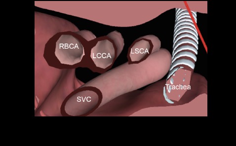
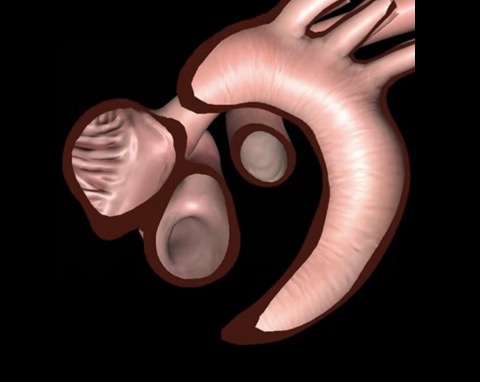
Abbreviations: 2C, two-chamber; AL, antero-lateral; ALC, anterolateral commissure; Alt, alternative; Ao, aorta; AoV, aortic valve; CWD, continuous-wave Doppler; IVS, interventricular septum; LAX, long-axis; LCCA, left common carotid artery; LSCA, left subclavian artery; LV, left ventricular; LVOTO, left ventricular outflow tract; MV, mitral valve; MPA, main pulmonary artery; PM, postero-medial; PMC, postero-medial commissure; PV, pulmonic valve; PWD, pulsed-wave Doppler; RA, right atrium; RBCA, right brachiocephalic artery; RPA, right pulmonary artery; RV, right ventricle; RVOT, right ventricular outflow tract; RVOTO, right ventricular outflow tract; SAM, systolic anterior motion; SAX, short-axis; SVC, superior vena cava; TV, tricuspid valve. Adapted from Rousou 48, Eltzschig 40, Reeves 73, Kumbarathi 45, Stern 74 and Royse et al. 70 |
|||||||||||
eTable 6.6 Suitable epicardial views for congenital lesions
| Pathology | View |
Atrial septal defect |
LAX, 4C |
Total anomalous pulmonary venous connection |
confluence in relation to LA LAX, 4C |
Cor triatriatum |
SAX |
Mitral valve anomalies |
LAX, 4C, SAX |
Subvalvular apparatus of mitral valve |
SAX |
Left ventricle function |
SAX |
Subvalvular and valvular aortic stenosis |
LAX |
Subvalvular and valvular pulmonary stenosis |
RVOT |
Branch pulmonary artery stenosis |
Trans-aortic SAX |
Coarctation |
Trans-aortic LAX |
Glenn shunt |
Trans-aortic LAX |
Abbreviations: 4C, four-chamber; LA, left atrium, LAX, long-axis; RVOT, right ventricular outflow tract; SAX, short-axis; Adapted from 53 |
|