Figures
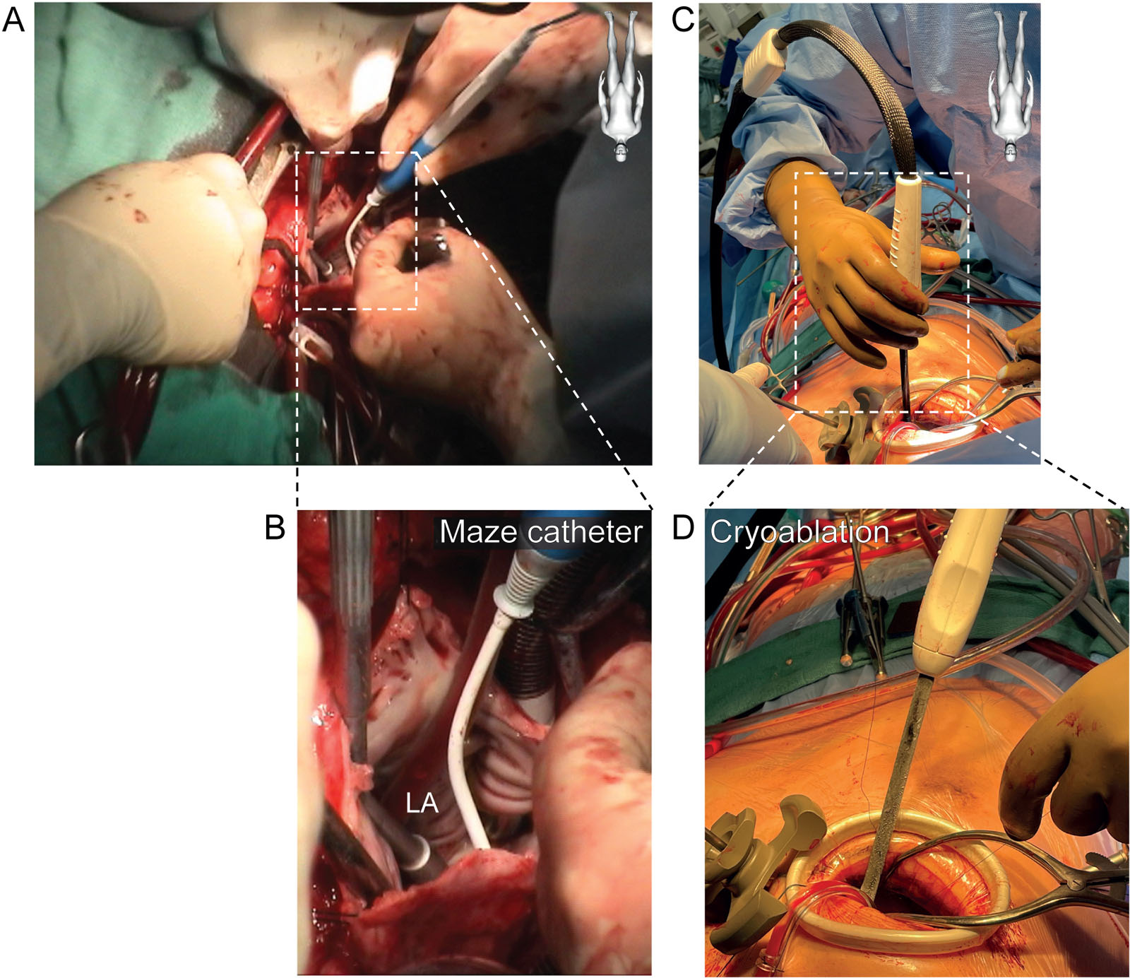
eFigure 8.2
Maze procedure (A,B) During the closure of an atrial septal defect in a 48-year-old man, the surgeon performs a Maze procedure for chronic atrial fibrillation. At that point, the echocardiographer pulls back the TEE probe to above the LA to avoid esophageal damage. (C,D) Cryoablation is applied around the pulmonary veins in order to prevent atrial fibrillation. Using this technique, there is no need of removing the TEE probe. Abbreviation: LA, left atrium; TEE, transesophageal echocardiography.
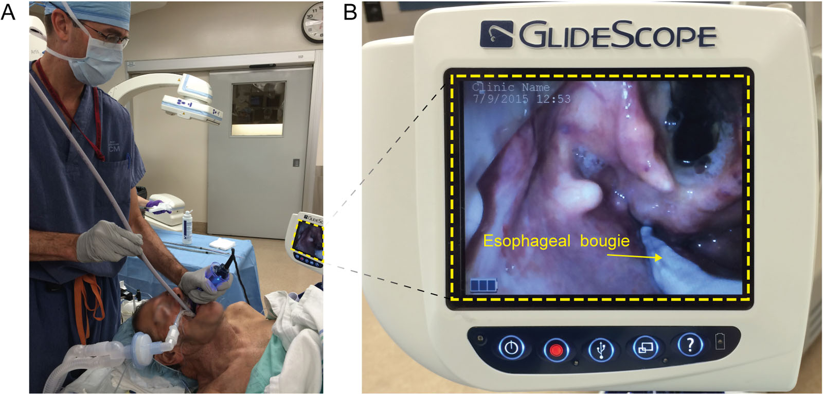
eFigure 8.5
Esophageal dilatation. (A,B) Progressive esophageal dilatation using a bougie under direct visualization in a patient with esophageal strictures to allow the insertion of a transesophageal echocardiography probe. (Curtesy of Dr Georges Desjardins)
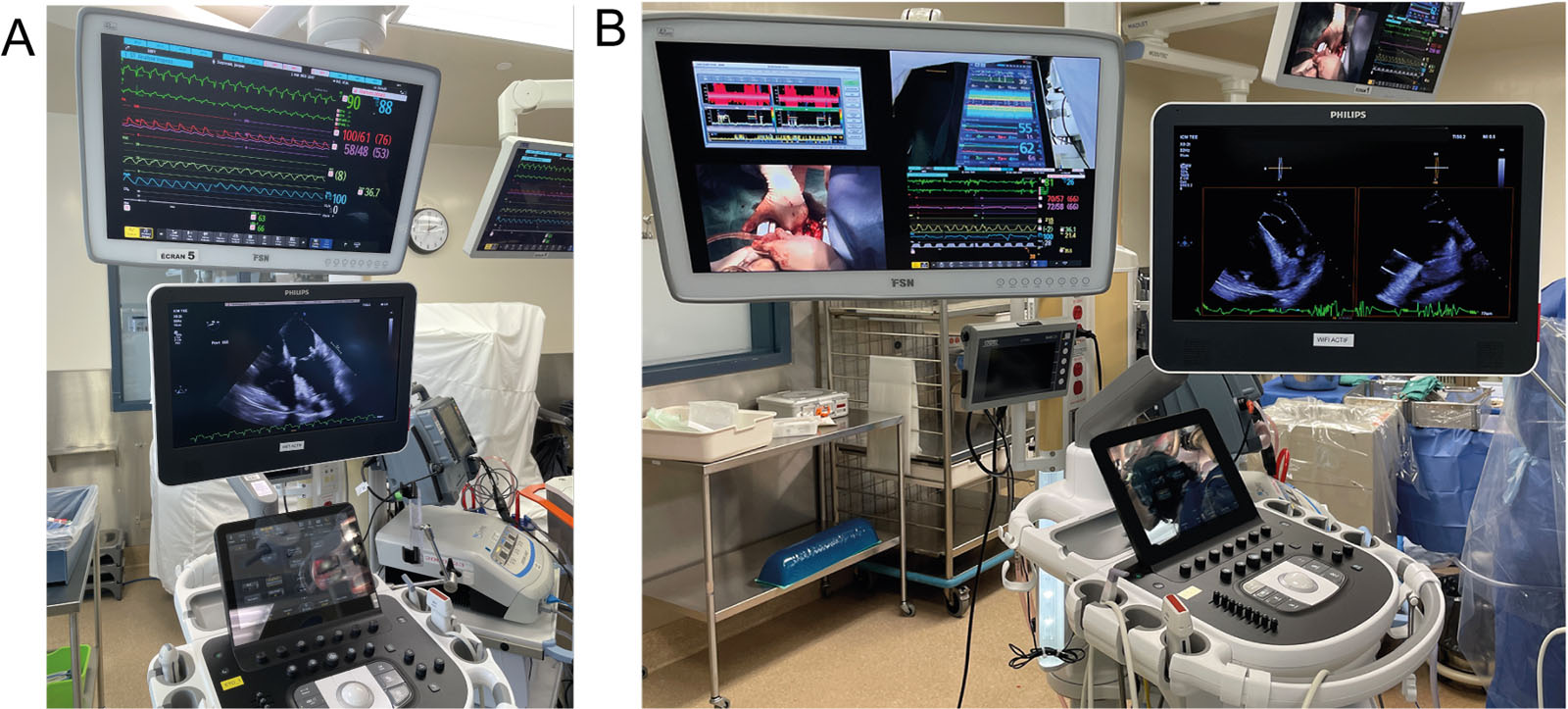
eFigure 8.6
Intraoperative displays. Intraoperative displays of both TEE imaging with (A) hemodynamic monitoring, (B) dual hemodynamic and intraoperative imaging, or (C) multimodal cerebral, hemodynamic and intraoperative monitoring. Abbreviations: TEE, transesophageal echocardiography.
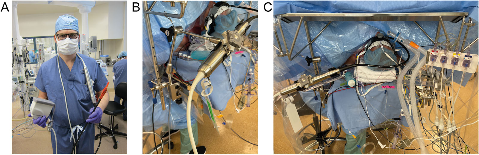
eFigure 8.14
TEE probe protection. (A) The operator holds the connector and distal probe in both hands during transport to avoid damaging the TEE probe. (B) Custom made TEE probe holder66 and (C) satellite operating metallic table can stabilize and protect the TEE probe. Abbreviation: TEE, transesophageal echocardiography.
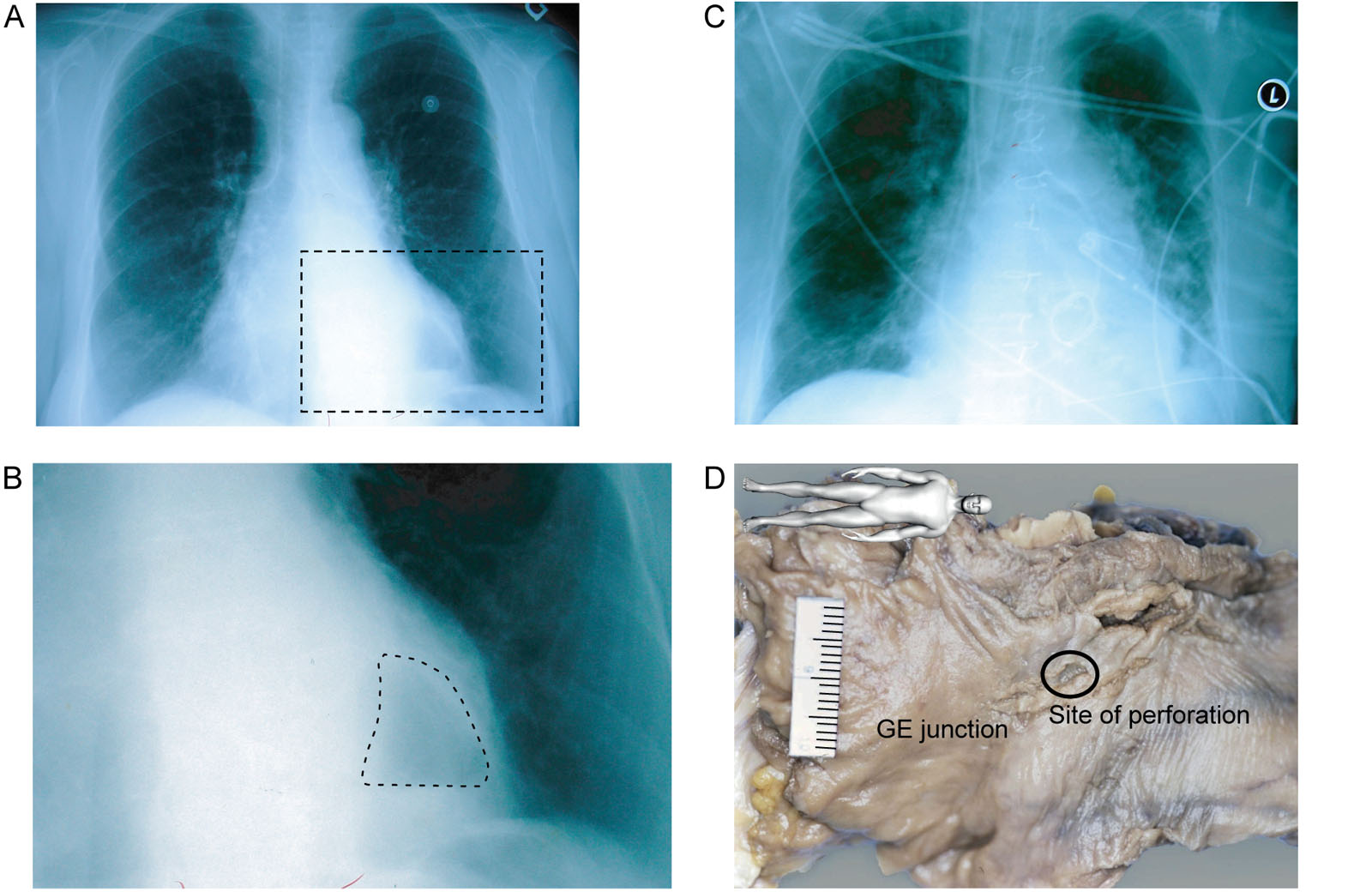
eFigure 8.17
Distal esophageal perforation. A 71-year-old woman undergoes AoV and MV replacement with TEE monitoring. (A,B) Her preoperative chest X-ray shows a hiatal hernia (dotted line). (C) In the postoperative period, she developed a left-sided pneumothorax and pleural effusion requiring a chest tube. She then went into multi-organ failure and died. (D) The autopsy showed distal esophageal perforation and a posterior mediastinal abscess. Abbreviations: AoV, aortic valve; GE, gastroesophageal; L, left; MV, mitral valve; TEE, transesophageal echocardiography. Source: Photo D courtesy of Dr. Tack Ki Leung.
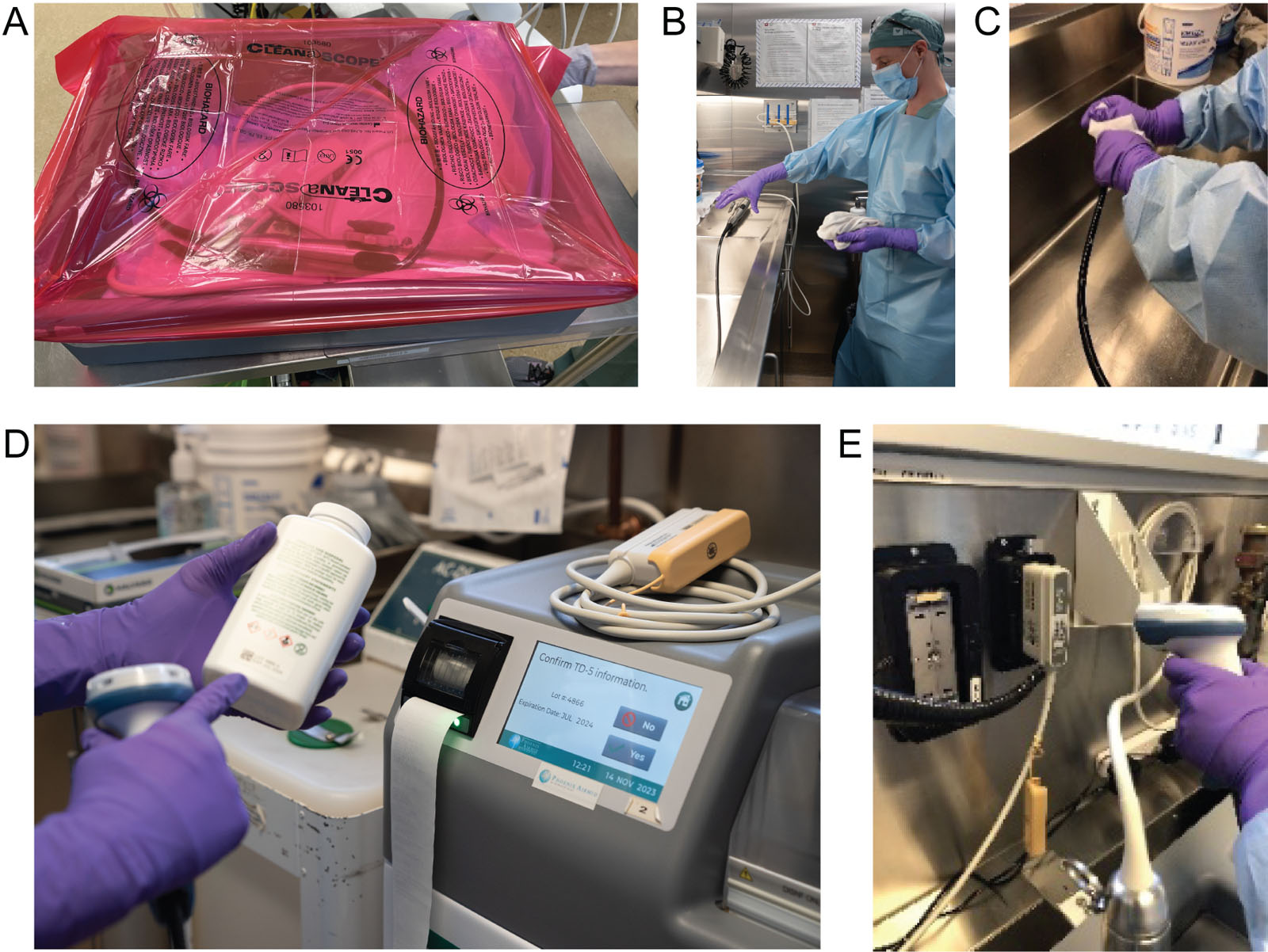
eFigure 8.22
MDRD. (A) The soiled TEE probe arrives in a red-covered plastic box in the MDRD. (B, C) The probe undergoes a manual inspection and washing using an enzymatic solution. (D) After probe transfer to an automated probe washer-disinfector (TEE-CLEAN using TD5) for a 30 minutes cycle followed by hand-held drying. (E) The disinfectant, cleaning enzyme, and the probe serial number are scanned for quality control. Abbreviations; MDRD, medical device reprocessing department; TEE, transesophageal echocardiography.
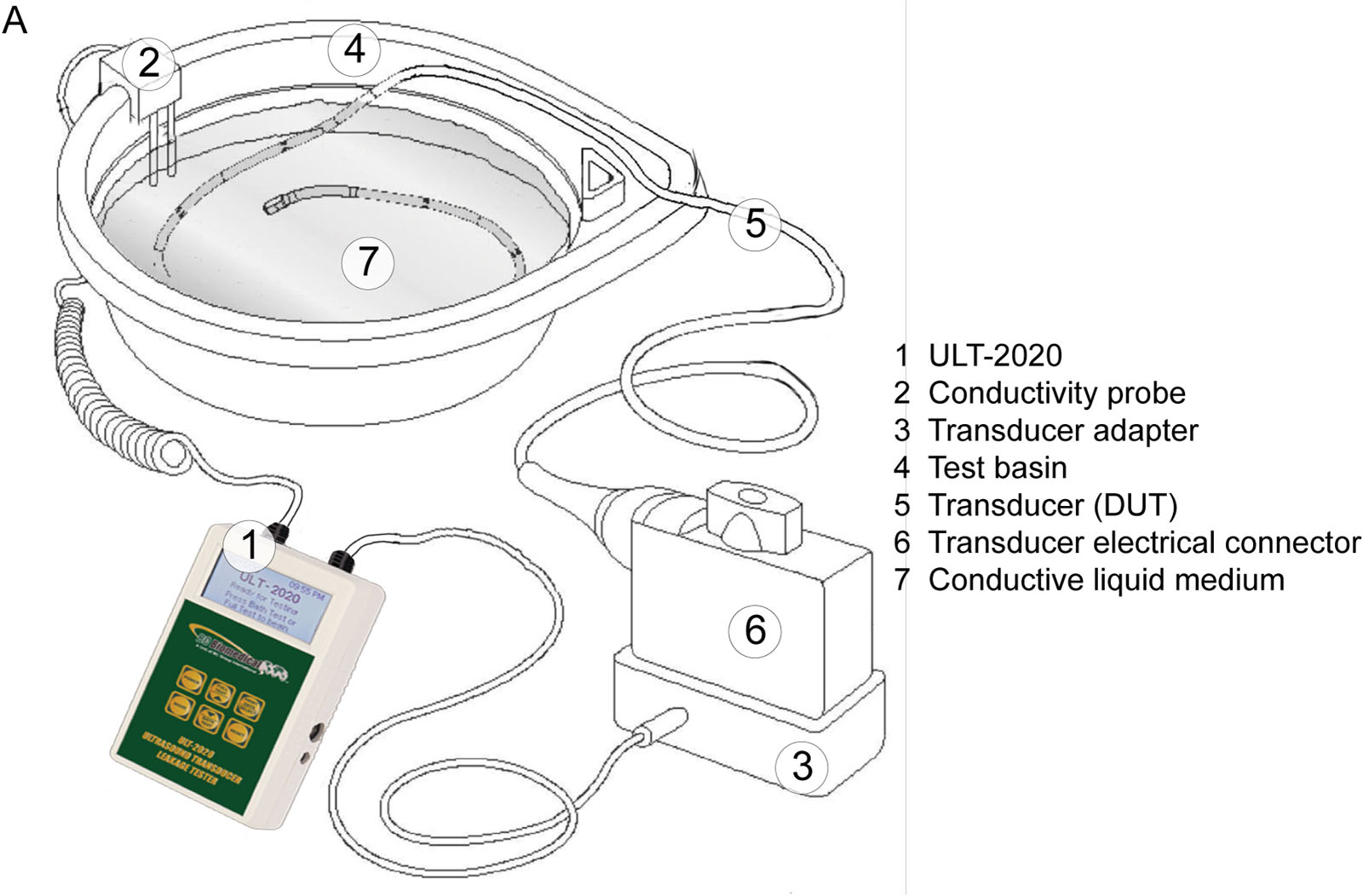
eFigure 8.23
Conductivity testing. This is the setup for ULT-2020, which is part of the routine verification for electrical leakage. Abbreviations; DUT, device under test. Adapted with permission of BC group St-Charles, MO 63301 USA.
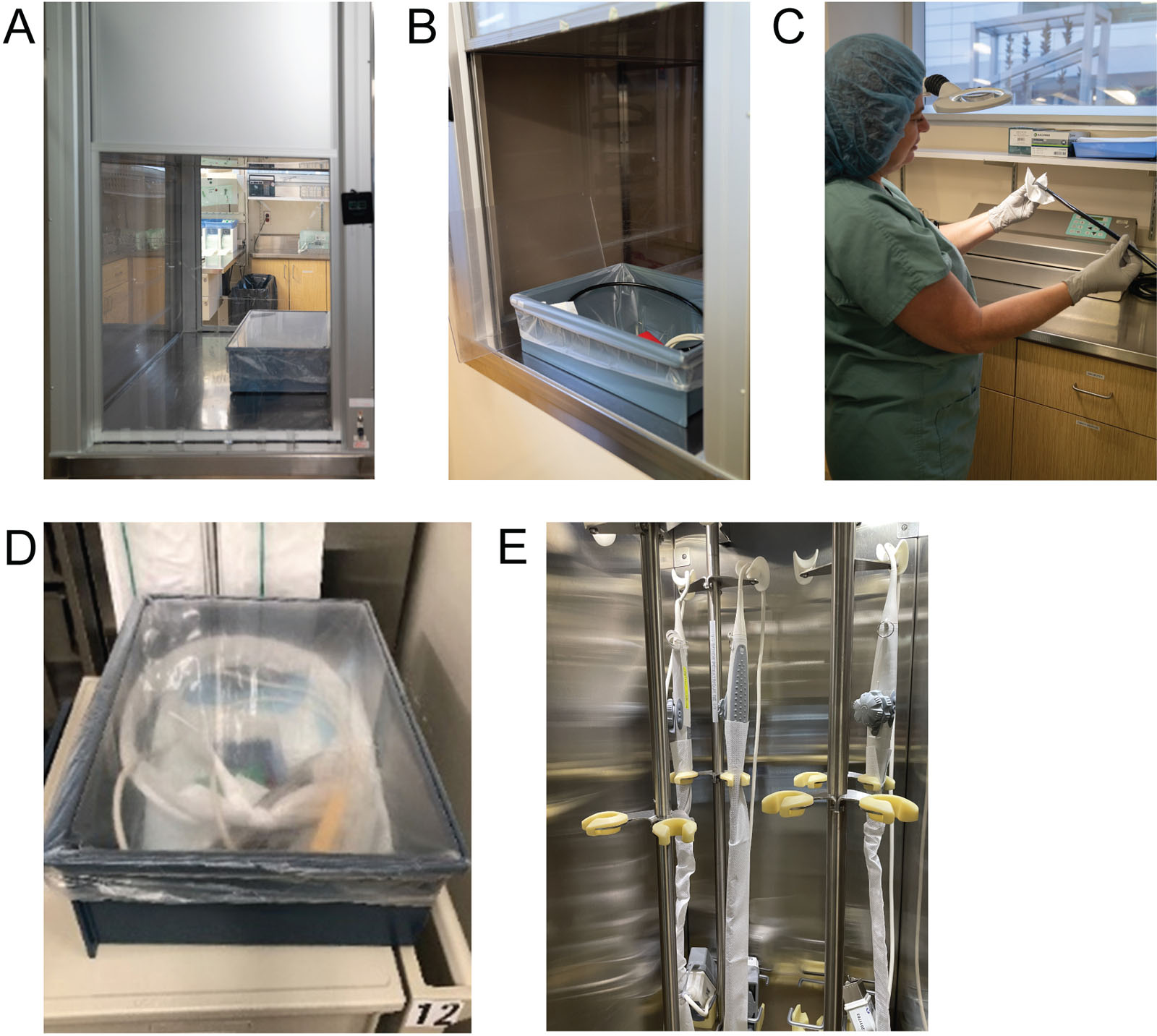
eFigure 8.24
Post-sterilization unit. (A) A pass-through hatch separates the soiled area from the clean area in the MDFR. (B) The clean TEE probe moves to this unit (C) for a second inspection and the attachment of a 7-day expiration tag. (D) The clean TEE probe is placed in a box with a clear plastic cover. After removing the probe from the cabinet, the enzyme foam is placed for future transport and post-op utilization. (F) In the OR, all clean TEE probes hang in ventilated storage cabinet. The entire cleaning process takes 90 minutes. Abbreviations: MDRD, medical device reprocessing department; OR, operating room; TEE, transesophageal echocardiography. Courtesy of Mounia Boulmerka.
Videos
Chapter 08 Fig01A
Chapter 08 Fig01B
Chapter 08 Fig04
Chapter 08 Fig04A
Chapter 08 Fig06A
Chapter 08 Fig06B
Chapter 08 Fig09A
Chapter 08 Fig09B
Chapter 08 Fig09C
Chapter 08 Fig09D
Chapter 08 Fig10AB
Chapter 08 fig10C
Chapter 08 Fig11A
Chapter 08 Fig11B
Chapter 08 Fig11C
Chapter 08 Fig11DEF
Chapter 08 Fig11G
Chapter 08 Fig20A
Chapter 08 Fig20B
Chapter 08 Fig20C
Chapter 08 Fig20DEF
Chapter 08 Fig21D
Chapter 08 Fig21E
Chapter 08 Fig22B
Chapter 08 Fig22D
Chapter 08 Fig22E
Chapter 08 Fig23A
Tables
eTable 8.4 Patient evaluation
|
Abbreviations: EKG, electrocardiogram; ETT, endotracheal tube; TEE, transesophageal echocardiography. |