Drag-and-Drop Self Tests
Instructions
Match the labels listed to the numbers as shown on the images to test your knowledge of key anatomical landmarks and structures. Simply drag the appropriate number to sit alongside the appropriate label, as shown above.
- Skull of full-term fetus – A from the front; B from above
- Skull of full-term fetus – C from the left
- Adult skull – from the right
- Adult skull with individual bones coloured – from the left
- Adult skull, median sagittal section hence removal of midline structures forming the nasal septum – from the left
- Adult skull, median sagittal section with removal of the nasal septum and individual bones coloured – from the right
- Cranial vault coverings – from above
- Brain, with arachnoid mater and underlying blood vessels removed from the right hemisphere – from above
- Brain, with arachnoid mater covering intact – from the left
- Brain, with arachnoid mater and underlying blood vessels removed – from the left
- Brain, median sagittal section, with arachnoid covering intact – from the right
- Brain, median sagittal section, with arachnoid and underlying blood vessels removed – from the right
- Dura mater and meningeal vessels – from the left
- Cranial cavity, paramedian sagittal section, hence the preservation of the falx cerebri and nasal septum – from the right
- Base of adult skull, external surface – from below
- Base of adult skull, internal surface – from above
- Brain, base, cerebellum and brainstem, with arachnoid mater and blood vessels removed from the left hemisphere – from below
- A. Cranial cavity, base – from above
- Cranial cavity, brain and upper spinal cord in a paramedian sagittal section, with removal of the falx cerebri and nasal septum – from the right
- Cranial cavity, brain and upper spinal cord in a paramedian sagittal section, with removal of the falx cerebri and nasal septum and exposure of cranial nerves in situ – from the right
- Cranial cavity, cavernous sinus, cranial nerves in situ – from the left above and slightly behind
- Cranial cavity, optic and olfactory nerves in situ – from above
- Lower brainstem and cervical part of the spinal cord – from behind
- Lower brainstem and cervical part of the spinal cord – from behind
- A. Superficial structures of the head, neck, shoulder and upper thorax – from the front and left
- Anterior cervical region (anterior triangle) of neck – from the front and left
- Lateral cervical region (posterior triangle) of neck – from the front, left and above
- A. Deep neck, great vessels – from the front
B. Isolated thyroid gland – from above
C. Isolated left superior parathyroid gland – from the right - A. Deep structures of neck I – from the left and slightly below
B. Isolated left submandibular gland – from above
C. Isolated left parotid gland – from above - Deep structures of neck II, with bisection and removal of the left half of pharynx and larynx – from the front left and slightly below
- Face, superficial structures I – from the left
- Face, superficial structures II – from the left
- Masticatory muscles I – from the left
- Masticatory muscles II, with masseter reflected inferiorly to display temporalis insertion – from the left
- Infratemporal fossa I, with partial reduction of the mandible and exposure of the mandibular canal – from the left
- Infratemporal fossa II, larynx and deep neck, with removal of the majority of the mandible – from the left
- Submandibular region and larynx – from the left
- A. Larynx – from the front
- B. Larynx – from behind
- Hyoid bone and cartilages of the larynx – A from the front; B from behind
- Pharynx posterior surface – from behind
- Adult skull, anterior external base, dentition – from below
- Adult mandible, dentition – from above
- A. Mouth and pharynx, paramedian sagittal section – from the right
B. Structures of the tongue exposed in a paramedian sagittal section – from the right
C. Isolated left sublingual gland – from the right - Floor of mouth, deep and adjacent structures exposed in a paramedian sagittal section – from the right
- A. Nasal septum, paramedian sagittal section – from the right
- B. With partial removal of the cribriform plate of the ethmoid bone and nasal septum to expose the mucosal lining – from the right
- A. Lateral wall of the nasal cavity and nasopharynx, paramedian sagittal section with removal of the nasal septum – from the right
- B. Semilunar hiatus, with extensive removal of the superior and middle nasal conchae – from the right
- A. Lateral wall of the nasal cavity, paramedian sagittal section with removal of the nasal septum and a portion of inferior nasal concha to display the opening of the nasolacrimal duct – from the right
- B. With removal of a posterior portion of the nasal conchae to expose the palatine canal – from the right
- Structures of the external nose – A, B from the left; C from the front
- Adult skull, structures relating to the orbit – from the front right and slightly above
- Adult skull, with coloured bones – from the front left and slightly above
- A. Left orbit with roof removed – from above
- B. Superficial structures – from above
- Left orbit with roof and lateral wall removed – from above left and slightly behind
- Left orbit with partial removal of roof and lateral wall – from the left
- Left orbit with partial removal of roof and lateral wall – from the left
- Left orbit with eyeball removed – from the front
- A. Left orbit and nasolacrimal duct – from the front and slightly left
B. Isolated left lacrimal gland – from above - A. Left auricle – from the left
- B. Left auricular cartilage (pinna) – from the left
- Adult skull without the mandible, lower lateral surface – from the left and slightly below
- Left inner ear exposed through the tegmen tympani of the temporal bone in the floor of the middle cranial fossa – from above
- Coronal section through the left ear – from behind
- Coronal section through the left ear – from the front
- Adult skeleton of the vertebral column – from the left
- Adult skeleton, with long bones of the left upper and lower limb removed – from the left
- A. Adult first (CI) cervical vertebra – atlas – from above
B. Adult second (CII) cervical vertebra – axis – from above
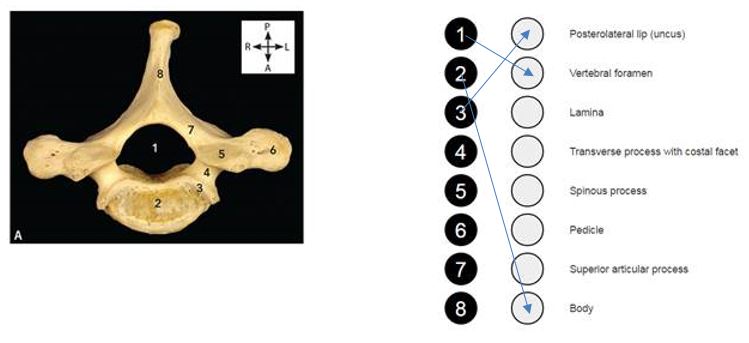
C. Adult fifth (CV) cervical vertebra – from above - A. Adult first (TI) thoracic vertebra – from above B. Adult fourth (LIV) lumbar vertebra – from above
- Skull and vertebral column, with posterior half of the cranium removed and vertebral canal opened to expose the brain and spinal cord in situ – from behind
- Skull and cervical part of the vertebral column, with the posterior part of the cranium removed, vertebral canal exposed and spinal cord partially removed – from behind
- Adult skeleton of the trunk – from the front
- Anterior muscles of the trunk – from the front
- Adult skeleton of the trunk – from behind
- Posterior muscles of the trunk – from behind
- Adult bones of the left upper limb – A from the front; B from behind
- Coronal section through the left shoulder joint – from the front
- Superficial structures of the left anterior thoracic wall and shoulder – from the front and slightly right
- Structures of the left deep lateral neck, brachial plexus, axilla, shoulder and upper arm – from the front
- Left brachial plexus and axilla I – from the front
- Left brachial plexus and axilla II – from the front
- Coronal section through the left elbow joint – from the front
- Adult bones of the left elbow joint – A from the front; B from behind
- Structures of the left anterior shoulder, upper arm and forearm – from the front
- Structures of the left posterior upper arm and forearm – from behind
- Superficial structures of the left forearm and palm of hand – from the front
- Superficial structures of the left distal forearm and dorsum of hand – from behind
- Adult bones of the left hand, palmar surface – from the front
- Adult bones of the left hand, dorsal surface – from behind
- Coronal section through the left wrist joint and hand – from behind
- Sagittal section through the joints of the left wrist and middle finger – from the left
- Superficial structures of the left forearm and palm of hand – from the front
- Deep structures of the left distal forearm and palm of hand – from the front
- Deep structures of the left forearm and palm of hand – from the front
- Deep structures of the left distal forearm and palm of hand – from the front
- Adult right first (I) atypical rib – A from above; B from below
- Adult right typical rib – A from above; B from below
- Adult skeleton of the thorax – from the front
- Muscles of the external thoracic wall – from the front
- Superficial structures of the female breast and external thoracic wall – from the front and left
- Sagittal section through the left female breast – from the left
- Thorax with ribcage and thoracic viscera in situ – from the front
- Thorax with ribcage removed and thoracic viscera in situ – from the front
- Right lung, lateral aspect – from the right
- Left lung, lateral aspect – from the right
- Right lung, medial aspect – from the left
- Right lung root and mediastinum – from the right
- Left lung root and mediastinum – from the left
- Left lung, medial aspect – from the right
- Heart – from the front
- Heart, superficial structures – from the front
- Heart – from behind
- Heart, superficial structures – from behind
- Heart, bisected by coronal section, anterior half – from behind
- Heart, bisected by coronal section, posterior half – from the front
- Heart, with right atrium exposed – from the front and slightly right
- Heart valves: pulmonary (open), aortic (closed) and mitral (closed), in situ – from above
- Diaphragm, superior surface (thoracic floor) – from above
- Diaphragm, inferior surface (abdominal roof) – from below
- Adult skeleton of the abdominal region – from the front
- Abdomen, muscles of the anterior wall – from the front
- Abdominal viscera I – from the front
Structures of the internal abdominal wall – from below - Abdominal viscera II, with greater omentum reflected superiorly – from the front
- Transverse section through the abdomen at the level of the second (LII) and third (LIII) lumbar vertebra – from below
- Caecum, terminal ileum (the iliocaecal junction) and vermiform appendix – from the front
- Transverse section through the abdomen at the level of the first (LI) lumbar vertebra – from below
- Upper abdominal viscera I, with removal of most of the small and all of the large intestine – from the front and slightly below
- Upper abdominal viscera II, with removal of most of the small and all of the large intestine – from the front and slightly below
- Stomach, incised along the length of the greater curvature and opened (as a book), to expose internal structures in a coronal plane, thus views are:
A. Anterior portion re ected superiorly, from behind
B. Posterior portion, from the front - Liver – from the front
- Liver – from below
- Pancreas – from the front
- Spleen – A. from the front
- Spleen – B. from below
- Posterior abdominal wall I – from the front
- Posterior abdominal wall II, with stomach and duodenum reflected superiorly – from the front
- Right kidney, with adrenal gland – from the front: A. Encapsulated within perinephric fat
- Right kidney, with adrenal gland – from the front: B. With perinephric fat removed and upper part within brous capsule
- Right kidney bisected by coronal section, posterior half – from the front
- Adult skeleton of pelvis, with ligaments – from the front
- Adult skeleton of pelvis, with ligaments – from above
- A. Structures within the male pelvis, left side in a paramedian sagittal section – from the right
B. Isolated left testicle – from the left - A. Structures within the female pelvis, left side in a paramedian sagittal section – from the right
B. Isolated left ovary – from the right - Male perineum, with body of penis in transverse section – from below
- Female perineum – from below
- Adult bones of the left lower limb – A from the front; B from behind
- Coronal section through the left hip joint – from the front
- Left gluteal region with gluteus maximus and medius severed and reflected laterally – from behind
- Right gluteal region with removal of the lower outer two-thirds of gluteus maximus – from behind
- Superficial structures of the left femoral triangle and thigh – from the front
- Superficial structures of the left gluteal region, thigh and popliteal fossa – from behind
- Superficial structures of the left leg – from the front
- Superficial structures of the left leg and popliteal fossa – from behind
- Sagittal section through the left knee joint – from the left
- Adult bones of the left knee joint – A from the front; B from behind
- Superficial structures of the left lower leg, ankle and dorsum of foot I – from the front left and slightly above
- Superficial structures of the left lower leg, ankle and dorsum of foot II – from the front left and slightly above
- Deep medial structures of the left lower leg, ankle and sole of foot I – from the right
- Deep medial structures of the left lower leg, ankle and second layer of sole of foot II – from the right and slightly below
- Adult bones of the left foot – from above
- Adult bones of the left foot – from below
- Coronal section through the left ankle joint and foot – from the front
- Sagittal section through the left ankle joint, foot and great toe – from the right
- Superficial structures of the left sole of foot – from below
- Structures of the first layer of the left sole of foot – from below